
 PDF Download
 PubReader
 Export Citation
 Email the Author
 Share Facebook
 Share Twitter
 Cited by 0 Article
 Metrics (View:12)








Introduction
Mango (Mangifera indica L.), a perennial evergreen tree belonging to the family Anacardiaceae, is one of the most economically important tropical fruits owing to its unique flavor, high nutritional value, and widespread consumer acceptance (Tharanathan et al., 2006; Kumar et al., 2021). In Korea, mango consumption has steadily increased with the growing demand for tropical fruits and processed mango products, including juice, puree, and dried snacks (Deeksha & Sunita, 2020; Korea Customs Service, 2024). Besides its desirable sensory properties, mango is an excellent source of carotenoids, vitamin C, and phenolic compounds such as mangiferin, quercetin, and gallic acid, which exhibit potent antioxidant and anti-inflammatory activities (Masibo & He, 2008; Kumar et al., 2021).
Recently, considerable attention has been directed toward mango processing by-products, particularly peel and seed, as sustainable sources of bioactive compounds. Mango seed, particularly the kernels contain abundant carbohydrates, proteins, lipids, and starch with favorable physicochemical and rheological properties, making them suitable for various food and industrial applications (Fariña et al., 2019; Mieles-Gómez et al., 2021). Likewise, mango peel contains considerable amounts of phenolic compounds and carotenoids, including mangiferin, quercetin, and ellagic acid, and has been reported to exhibit strong antioxidant activity and various biological functions (Manzoor et al., 2022; Kittiphoom, 2012). The composition of these bioactive compounds varies depending on cultivar, maturity stage, and environmental conditions (Trong et al., 2020).
Despite their nutritional and functional value, mango peel and seed account for approximately 15–20% and 10–25% of the total fruit weight, respectively, and are generally discarded during industrial processing, resulting in environmental burdens and the loss of valuable biomass (Naeem et al., 2019; Kaur et al., 2023). Consequently, the effective utilization of mango by-products has become increasingly important within the framework of sustainable food production, up-cycling, and zero-waste strategies (Kaur et al., 2023).
The recovery of bioactive compounds from plant materials is strongly influenced by extraction conditions, particularly solvent composition. Because phenolic compounds possess different polarities, extraction efficiency largely depends on the polarity of the extraction solvent (Naczk & Shahidi, 2004; Shi et al., 2005). Distilled water is a safe and environmentally friendly solvent but has limited ability to extract relatively hydrophobic phenolic compounds, whereas aqueous ethanol mixtures provide suitable polarity for recovering a broad range of phenolic constituents (Wang & Weller, 2006; de Camargo et al., 2016). Previous studies have demonstrated that ethanol concentration markedly influences phenolic recovery and antioxidant activity in plant-derived materials.
Ultrasound-assisted extraction (UAE) has recently emerged as an efficient and environmentally friendly extraction technology for improving the recovery of bioactive compounds. Cavitation generated during ultrasound treatment disrupts plant cell walls, enhances solvent penetration, and accelerates mass transfer, thereby facilitating the release of intracellular phytochemicals (Chemat et al., 2017). Furthermore, UAE enables efficient extraction under relatively mild conditions, minimizing the degradation of heat-sensitive compounds (Al-Dhabi et al., 2017). Numerous studies have demonstrated that UAE improves the extraction efficiency of phenolic compounds and antioxidant activities from plant materials and food by-products (Chemat et al., 2017).
Although the antioxidant potential of mango peel and seed has been reported previously, most studies have focused on individual by- product fractions or a single extraction condition. Differences in extraction solvent and extraction technique often make direct comparison among studies difficult. Consequently, comprehensive information comparing edible and non-edible mango tissues under different solvent systems together with ultrasound-assisted extraction remains limited. Such comparative information is essential for establishing practical extraction strategies and maximizing the utilization of mango processing by-products as value-added food resources.
Therefore, the present study compared the proximate composition, physicochemical properties, and color characteristics of mango pulp, peel, and seed obtained from Thai mangoes distributed in the Korean market. Furthermore, the effects of extraction solvents (distilled water, 70% ethanol, and 95% ethanol) and ultrasound-assisted extraction on total polyphenol and flavonoid contents as well as antioxidant activities were investigated. The findings of this study provide fundamental information for optimizing extraction conditions and support the sustainable utilization of mango by-products as natural antioxidant sources and value-added functional food ingredients.
Materials and Methods
Fresh Thai mangoes (Mangifera indica L.) imported into Korea were purchased through an online retail market and used as the experimental materials. Folin–Ciocalteu phenol reagent, (+)-catechin, gallic acid, and potassium persulfate were obtained from Sigma-ldrich Co. (St. Louis, MO, USA). All other chemicals and organic solvents used for proximate composition and antioxidant analyses were of analytical grade or higher and purchased from Junsei Chemical Co., Ltd. (Tokyo, Japan).
The mangoes were thoroughly washed under running water and surface moisture was removed using paper towels. Mango fruits were manually separated into peel, pulp, and seed. In this study, the entire seed (including the endocarp and kernel) was used for all analyses. Fresh samples were immediately used for the determination of proximate composition, pH, total acidity, soluble solids (°Brix), and color values. For antioxidant and bioactive compound analyses, each sample was frozen at –80˚C and subsequently freeze-dried using a freeze dryer (FDU-1200, EYELA, Tokyo, Japan) at a chamber pressure of 40 Pa for 48 h. The freeze-dried samples were ground using a grinder (PGR 002M, Supreme Electric Co., Ltd., Goyang, Korea) and passed through a 20-mesh sieve to obtain a homogeneous powder. The powdered samples were sealed in airtight.
The proximate composition of fresh mango pulp, peel, and seed was determined according to the official methods of the Association of Official Analytical Chemists (AOAC, 2005). All results are expressed on a fresh-weight basis. Moisture content was determined by the air-oven drying method at 105˚C. Crude protein content was determined using the semi-micro Kjeldahl method following acid digestion with sulfuric acid in the presence of a selenium catalyst (420˚C for 50 min), and the nitrogen content was quantified by automated titration. Crude fat content was determined by ether extraction using a Soxtec system (Soxtec System HT 1043, Foss Tecator, Eden Prairie, MN, USA). Ash content was determined gravimetrically after incineration in a muffle furnace at 550˚C. Crude fiber content was determined using a Fibertec system after sequential acid and alkali digestion according to the AOAC method.
Fresh mango pulp, peel, and seed (200 g each) were homogenized with an equal volume (w/v) of distilled water using a homogenizer and centrifuged. The supernatant was filtered through Whatman No. 1 filter paper (Cytiva, Marlborough, MA, USA) and used for subsequent analyses.
The pH was measured using a pH meter (SevenCompact™ S220, Mettler-Toledo, Zurich, Switzerland). Soluble solids content was determined using a digital refractometer (PR-201α, Atago Co., Tokyo, Japan) and expressed as °Brix. Total acidity was determined by titrating 10 mL of the filtrate with 0.1 N NaOH to an endpoint of pH 8.3 and expressed as percentage (%) citric acid equivalents.
The surface color of fresh mango pulp, peel, and seed was measured using a Chroma Meter (CR-400, Konica Minolta, Tokyo, Japan). The instrument was calibrated using a standard white calibration plate (L*=97.10, a*=0.24, and b*=1.75) before measurement. Color values were expressed according to the CIE L*, a*, and b* color system.
Fresh mangoes were washed thoroughly with tap water and separated into pulp, peel, and seed. Each fraction was frozen at –80˚C and freeze-dried using a freeze dryer (FDU-1200, EYELA, Tokyo, Japan). The dried samples were ground using a grinder (PGR 002M, Supreme Electric Co., Ltd., Goyang, Korea) and passed through a 20-mesh sieve to obtain homogeneous powders.
To evaluate the effect of extraction solvents, powdered samples (10 g) were mixed with 20 mL of distilled water, 70% ethanol, or 95% ethanol (solid-to-solvent ratio, 1:2, w/v). These solvents were selected because they are commonly used for the extraction of plant phenolic compounds and represent a wide range of solvent polarities, including water, aqueous ethanol, and nearly absolute ethanol. The mixtures were homogenized and extracted at room temperature for 30 min using an orbital shaker (BF-500SK, Biofree, Seoul, Korea). After extraction and centrifugation, the collected supernatants were diluted 1:4 (v/v) with the corresponding extraction solvent (5-fold dilution). The diluted extracts were used to determine TPC, TFC, DPPH and ABTS radical scavenging activities, and reducing power. The same dilution factor was applied to all samples to ensure consistent assay conditions. To compare extraction methods, 95% ethanol was selected based on the results of the solvent comparison experiment. Although 70% ethanol yielded higher total polyphenol and total flavonoid contents, 95% ethanol exhibited the highest overall antioxidant activities, including DPPH radical scavenging activity, ABTS radical scavenging activity, and reducing power. Therefore, 95% ethanol was selected for subsequent extraction method comparison. Conventional extraction was performed by incubating the sample–solvent mixture at room temperature for 30 min without ultrasound treatment. Ultrasound-assisted extraction was conducted for 30 min using a probe-type ultrasonic processor (VCX 750, Sonics & Materials Inc., Newtown, CT, USA) equipped with a temperature sensor and soundproof chamber. The ultrasound was applied at a frequency of 20 kHz and an amplitude of 50% under continuous mode. An ice-water bath was used during sonication to minimize heat generation and maintain the sample temperature at approximately 30˚C.
Following extraction, all samples were centrifuged at 13,500×g for 15 min, and the supernatants were filtered through Whatman No. 1 filter paper. The filtrates were appropriately diluted and used for the determination of total polyphenol content, total flavonoid content, DPPH and ABTS radical scavenging activities, and reducing power.
Total polyphenol content (TPC) was determined using the Folin–Ciocalteu method with slight modifications from the procedure described by Singleton & Rossi (1965). Briefly, 100 μL of the sample extract was mixed with 100 μL of 2 N Folin–Ciocalteu reagent and allowed to react for 3 min. Subsequently, 400 μL of 2% sodium carbonate (Na2CO3) solution was added, and the mixture was incubated in the dark for 1 h at room temperature. The absorbance was measured at 760 nm using a microplate reader, and the results were expressed as micrograms of gallic acid equivalents per gram of sample (μg GAE/g) based on a gallic acid standard curve.
Total flavonoid content (TFC) was determined according to the method of Zhishen et al. (1999). The diluted extract was mixed with an equal volume of 2% aluminum chloride (AlCl3) solution and incubated at room temperature for 15 min. The absorbance was measured at 430 nm, and the results were expressed as micrograms of catechin equivalents per gram of sample (μg CE/g) using a catechin standard curve.
DPPH radical scavenging activity was determined according to the method of Cheung et al. (2003) with slight modifications. Briefly, 0.2 mM DPPH solution was mixed with an equal volume of the sample extract and incubated in the dark at 37˚C for 30 min. The absorbance was measured at 515 nm, and the radical scavenging activity was expressed as percentage inhibition relative to the control.
ABTS radical scavenging activity was determined according to the method of Re et al. (1999). The ABTS radical cation (ABTSo+) was generated by mixing 7.0 mM ABTS with 2.45 mM potassium persulfate and allowing the mixture to stand in the dark at room temperature for 24 h. The resulting solution was diluted with ethanol to an absorbance of 0.73±0.03 at 735 nm. The sample extract was mixed with the ABTS working solution and incubated at 37˚C for 30 min. The absorbance was measured at 735 nm, and the radical scavenging activity was expressed as percentage inhibition.
Reducing power was determined according to the method of Oyaizu (1986). Briefly, 1 mL of the sample extract was mixed with 1 mL of 200 mM phosphate buffer (pH 6.6) and 1 mL of 1% potassium ferricyanide, followed by incubation at 50˚C for 20 min. After the addition of 10% trichloroacetic acid (TCA), the mixture was centrifuged, and 1 mL of the supernatant was mixed with distilled water and ferric chloride (FeCl3) solution. The absorbance was measured at 700 nm, and higher absorbance values indicated greater reducing power.
All experiments were performed in triplicate, and the results are expressed as the mean±standard deviation. Statistical analyses were performed using R software (version 3.5.1; R Foundation for Statistical Computing, Vienna, Austria). One-way analysis of variance (ANOVA) was used to evaluate differences among treatments, followed by Duncan’s multiple range test for post hoc comparisons. Differences were considered statistically significant at p<0.05.
Results and Discussion
The proximate composition of mango pulp, peel and seed is presented in Table 1. On a fresh-weight basis, the peel and seed exhibited higher protein, fat, and ash contents than the pulp. These differences may be partially attributed to the lower moisture contents of the peel and seed compared with the pulp. The moisture content was highest in the pulp (82.64%), followed by the peel (76.94%) and the seed (66.46%). This difference is attributed to the physiological characteristics of each tissue, as the pulp functions as a succulent edible tissue with a high water-holding capacity, whereas the seed forms a rigid, lignified structure containing less moisture.
The crude protein content was significantly higher in the by-products, with the peel (0.76%) and seed (0.62%) containing 1.58- and 1.29-fold higher protein levels, respectively, than the pulp (0.48%). Likewise, crude fat content followed the order of peel (0.26%)>seed (0.17%)>pulp (0.03%). These results suggest that, on a fresh-weight basis, mango peel and seed exhibited higher protein and fat contents than the edible pulp, although these differences may be partially influenced by differences in moisture content among the tissues. Similar trends have been reported by Ajila & Prasada Rao (2013) and Kim (2017), who demonstrated that mango peel and seed are valuable sources of proteins and lipids rather than merely processing residues. The present findings further support the potential utilization of these by-products as nutritional ingredients for value-added food applications.
The crude ash content was lowest in the pulp (0.16%), whereas the peel and seed contained 0.44% and 0.61%, respectively, indicating considerably higher mineral contents in the by-product fractions. In particular, the seed exhibited the highest crude fiber content (14.92%), followed by the peel (2.64%) and pulp (0.71%). The crude fiber content of the seed was more than 20-fold higher than that of the pulp, reflecting its structural role in protecting the embryo and supporting seed development. The high crude fiber content observed in the seed is consistent with its structural function. Previous studies have reported high dietary fiber contents in mango seeds and peels (Ajila et al., 2007; Ajila & Prasada Rao, 2013); however, because only crude fiber was determined in the present study, direct comparisons with total dietary fiber should be made with caution. Further evaluation of total dietary fiber would help clarify the potential application of mango seed as a dietary fiber ingredient in food products.
Overall, the proximate composition differed considerably among mango tissues, demonstrating that each fraction possesses distinct nutritional characteristics. While the pulp was characterized by its high moisture content, the peel contained relatively higher protein and fat contents, whereas the seed exhibited the highest crude fiber and mineral contents. These compositional differences indicate that mango processing by-products possess distinct compositional characteristics and may have potential for food applications, depending on their composition.
The pH, total acidity, and soluble solids content (SSC) of the mango pulp, peel, and seed are presented in Table 2. The SSC was significantly highest in the pulp (1.85 °Brix), followed by the seed (1.30 °Brix) and peel (0.95 °Brix) (p<0.05). As the primary edible portion of the fruit, the pulp functions as the principal storage tissue for water-soluble constituents during fruit development (Silva et al., 2008). Accordingly, the pulp exhibited a higher soluble solids content than the peel and seed. However, because SSC measured by a refractometer represents total soluble solids rather than individual sugars, the higher SSC observed in the pulp should not be interpreted as a direct measure of sugar concentration. Previous studies have reported that soluble sugars increase during mango fruit ripening through the conversion of starch into low-molecular-weight sugars (Trong et al., 2020); however, individual sugars were not determined in the present study.
| Measurement | Fruit part | ||
|---|---|---|---|
| Pulp | Peel | Seed | |
| pH | 4.02±0.01c | 4.48±0.01a | 4.43±0.00b |
| Total acidity (%) | 0.21±0.04a | 0.12±0.00b | 0.15±0.00ab |
| Soluble solids content (°Brix) | 1.85±0.07a1) | 0.95±0.07c | 1.30±0.00b |
The pH values of the peel (4.48) and seed (4.43) were slightly higher than that of the pulp (4.02), whereas total acidity was highest in the pulp (0.21%), followed by the seed (0.15%) and peel (0.12%). These results indicate that organic acids are more concentrated in the edible pulp than in the by-product fractions. Organic acids, including citric and malic acids, play important roles in determining fruit flavor, maintaining cellular metabolism, and contributing to microbial stability during postharvest storage (Zhang et al., 2023). Therefore, the lower pH and higher acidity observed in the pulp are likely associated with the characteristic sweet–acid balance of ripe mango fruit. In contrast, the relatively lower acidity of the peel and seed suggests compositional differences among mango tissues, which may influence their physicochemical properties and subsequent processing characteristics.
Overall, the significant differences in soluble solids, pH, and total acidity among mango tissues reflect their distinct physiological functions and compositional characteristics. These findings provide fundamental information for understanding the quality characteristics of mango by-products and their potential utilization as food materials.
The color characteristics of different mango parts are summarized in Table 3. The lightness (L*) was significantly highest in the pulp (46.75), followed by the peel (44.61) and the seed (39.53) (p<0.05), indicating that the edible pulp exhibited a brighter appearance than the by-product fractions. Redness (a*) was highest in the peel (3.82), whereas the pulp (2.84) and seed (1.88) showed significantly lower values. Regarding yellowness (b*), the pulp (15.26) and peel (14.89) exhibited similar values, while the seed showed a significantly lower value (9.41).
| Measurement | Fruit part | ||
|---|---|---|---|
| Pulp | Peel | Seed | |
| Lightness (L*) | 46.75±0.27a1) | 44.61±0.35b | 39.53±0.16c |
| Redness (a*) | 2.84±0.02b | 3.82±0.07a | 1.88±0.05c |
| Yellowness (b*) | 15.26±0.16a | 14.89±0.32a | 9.41±0.03b |
The relatively high L* and b* values observed in the pulp are consistent with the accumulation of carotenoid pigments during fruit ripening (Sivankalyani et al., 2016). In particular, β-carotene is known to be one of the major pigments responsible for the characteristic yellow color of ripe mango and has been widely used as an indicator of fruit maturity and quality (Trong et al., 2020). The higher a* value observed in the peel is likely associated with chlorophyll degradation accompanied by the accumulation of anthocyanins and red-colored carotenoids during ripening (Sivankalyani et al., 2016). In contrast, the relatively low L* and b* values of the seed may be associated with its compact internal structure and other physical characteristics that influence light reflectance contribute to its darker appearance.
Overall, each mango fraction exhibited distinct color characteristics that reflect differences in tissue properties. Although the pulp and peel showed similar b values, these results should not be interpreted as indicating similar pigment contents. The observed color characteristics are generally consistent with previous reports describing mango tissues (Sivankalyani et al., 2016; Trong et al., 2020). In addition, previous studies have reported that mango peel contains various naturally occurring pigments and bioactive compounds (Ajila et al., 2007; Trong et al., 2020), although pigment composition was not determined in the present study.
TPC and TFC of mango pulp, peel, and seed extracted with different solvents are presented in Fig. 1. Among the extraction solvents evaluated, 70% ethanol consistently yielded the highest TPC and TFC in all mango tissues, with significantly higher values observed in the peel and seed than in the pulp (p<0.05). In particular, the seed extracted with 70% ethanol exhibited the highest TPC (387.71 μg GAE/g) and TFC (201.85 μg CE/g). These findings indicate that the non-edible portions of mango, especially the peel and seed, contained higher levels of phenolic compounds than the edible pulp under the extraction conditions evaluated.
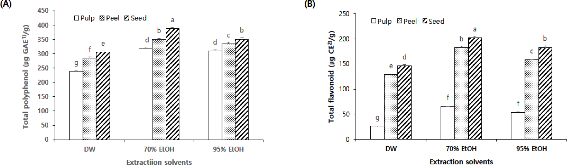
The higher TPC and TFC observed in mango peel and seed are consistent with previous studies reporting that these tissues contain abundant phenolic compounds associated with plant defense mechanisms (Ramírez-Brewer et al., 2025). Likewise, previous studies have suggested that the relatively high flavonoid content of mango seed may be associated with protection against oxidative stress during seed development (Manzoor et al., 2022; Choudhary et al., 2026). Previous studies have likewise reported that mango by-products contain abundant phenolic constituents, including mangiferin, a characteristic xanthone with well-documented antioxidant and anti-inflammatory activities (Masibo & He, 2008). Although individual phenolic compounds were not identified in the present study, the higher TPC and TFC observed in the peel and seed are consistent with these previous findings.
Extraction efficiency was markedly influenced by solvent polarity. Compared with distilled water and 95% ethanol, 70% ethanol consistently produced higher TPC and TFC values across all mango fractions. This finding is consistent with previous studies demonstrating that aqueous ethanol mixtures provide an optimal polarity for extracting phenolic compounds from plant materials (Barrales et al., 2018; Ferreira et al., 2023).
The effects of UAE on the TPC and TFC of mango pulp, peel, and seed are presented in Fig. 2. Because 95% ethanol consistently exhibited superior overall antioxidant activities in the solvent comparison experiment, despite the higher TPC and TFC obtained with 70% ethanol, it was selected for the subsequent comparison of conventional and ultrasound-assisted extraction methods. The solvent comparison and extraction-method comparison were conducted as independent experiments with different objectives; therefore, the results were interpreted within each experimental set rather than by direct comparison of absolute values between experiments. UAE significantly increased both TPC and TFC in all mango tissues. TPC increased by approximately 1.80-, 1.13-, and 1.12-fold in the pulp, peel, and seed, respectively, while TFC increased by approximately 1.72-, 1.22-, and 1.26-fold. Interestingly, the pulp exhibited the greatest relative increase despite having the lowest initial phenolic content, suggesting that ultrasound efficiency depends not only on phenolic concentration but also on tissue structure and the accessibility of intracellular compounds. The softer parenchymatous tissue of the pulp may be more susceptible to cavitation-induced disruption than the more rigid peel and seed tissues, resulting in greater relative release of phenolic compounds during extraction.
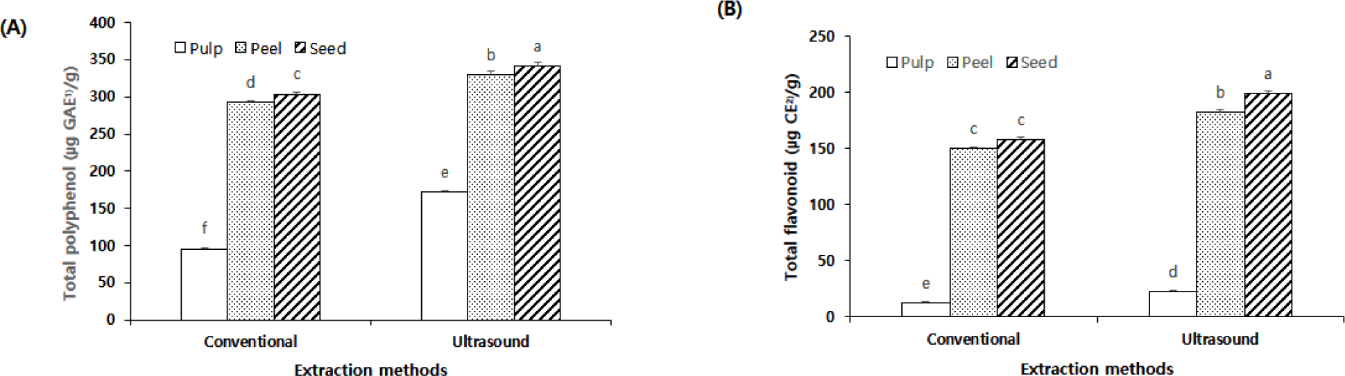
The enhanced recovery of phenolic compounds by UAE is attributed to cavitation, which disrupts plant cell walls, improves solvent penetration, and accelerates mass transfer (Chemat et al., 2017). In addition, UAE minimizes thermal degradation of heat-ensitive phytochemicals under relatively mild extraction conditions (Al-Dhabi et al., 2017). Although the relative enhancement induced by UAE was smaller in the peel and seed, these tissues maintained substantially higher absolute TPC and TFC values than the pulp after extraction.
Overall, the present results demonstrate that both extraction solvent and UAE markedly influenced the recovery of phenolic compounds from mango tissues. The consistently higher phenolic contents observed in the peel and seed further support their potential utilization as promising sources of phenolic-rich functional ingredients.
The antioxidant activities of mango pulp, peel, and seed extracted with different solvents are presented in Fig. 3. Overall, antioxidant activities varied depending on the mango tissue and extraction solvent. In general, the peel and seed exhibited higher antioxidant activities than the pulp, although not all comparisons were statistically significant. Under the extraction conditions evaluated, the highest DPPH radical scavenging activities were observed in the peel (76.30%) and seed (76.18%) extracted with 95% ethanol, whereas the pulp exhibited significantly lower activity. Likewise, the highest ABTS radical scavenging activities were obtained from the peel (71.75%) and seed (71.02%), whereas the greatest reducing power was observed in the seed (1.30). These results suggest that mango peel and seed exhibited higher antioxidant activities than the pulp under the extraction conditions evaluated.
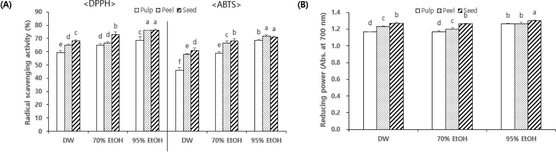
The antioxidant activity patterns were generally consistent with the TPC and TFC results, suggesting that phenolic compounds may contribute to the free radical scavenging and electron-donating capacities of mango tissues (Safdar et al., 2022). Previous studies have likewise reported that mango peel and seed contain abundant antioxidant phytochemicals, including mangiferin, gallic acid, and flavonoids, which contribute substantially to their antioxidant properties (Masibo & He, 2008; Manzoor et al., 2022).
Extraction solvent markedly influenced antioxidant activity. Although 70% ethanol yielded the highest total phenolic and flavonoid contents, the strongest radical scavenging activities were observed in the 95% ethanol extracts. This finding suggests that antioxidant activity may be influenced not only by the total phenolic and flavonoid contents but also by the composition and reactivity of the extracted compounds (Oubannin et al,, 2024). However, the specific compounds responsible for the higher antioxidant activity of the 95% ethanol extracts were not identified in the present study. Similar findings have been reported for mango by-products and other plant materials, where ethanol extracts exhibited stronger antioxidant activities than aqueous extracts (Ajila et al., 2007; Barrales et al., 2018).
The effects of UAE on the antioxidant activities of mango pulp, peel, and seed are presented in Fig. 4. Because the solvent comparison and UAE experiments were conducted independently under different analytical conditions, the results were interpreted within each experimental set rather than by direct comparison of absolute values between experiments.
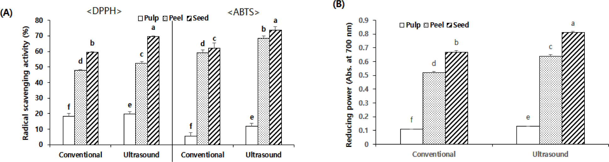
UAE further enhanced antioxidant activities in all mango tissues. DPPH radical scavenging activity increased from 18.28% to 19.66% in the pulp, from 47.71% to 52.42% in the peel, and from 59.41% to 60.66% in the seed. Likewise, ABTS radical scavenging activity increased from 5.74% to 11.81% in the pulp, from 75.16% to 76.45% in the peel, and from 72.25% to 74.81% in the seed, whereas reducing power increased from 0.11 to 0.13 in the pulp, from 0.52 to 0.64 in the peel, and from 0.67 to 0.81 in the seed. Among the three tissues, the pulp exhibited the greatest relative improvement following UAE, particularly in ABTS radical scavenging activity, despite its comparatively low initial antioxidant capacity. This finding suggests that ultrasound may facilitate the release of intracellular antioxidant compounds more effectively from the softer pulp tissue than from the structurally rigid peel and seed.
The enhancement of antioxidant activity by UAE is attributed to cavitation-induced disruption of plant cell walls, which improves solvent penetration and accelerates mass transfer during extraction (Chemat et al., 2017). Moreover, UAE minimizes thermal degradation of heat-sensitive phytochemicals under relatively mild extraction conditions (Al-Dhabi et al., 2017). Although the relative increase induced by UAE was greater in the pulp, the peel and seed consistently maintained superior absolute antioxidant activities, indicating that these by-products remain the most promising sources of natural antioxidants.
Overall, the present results demonstrate that both extraction solvent and UAE substantially influenced the antioxidant activities of mango tissues. While 70% ethanol was more effective for recovering total phenolics and flavonoids, 95% ethanol generally produced stronger radical scavenging activities, suggesting that antioxidant capacity depends not only on the total amount of phenolic compounds but also on differences in their composition and antioxidant potency. The consistently higher antioxidant activities observed in the peel and seed further support their potential utilization as sustainable sources of natural antioxidants and value-added functional food ingredients.
Conclusion
This study investigated the proximate composition, physicochemical properties, color characteristics, bioactive compounds, and antioxidant activities of mango pulp, peel, and seed, together with the effects of extraction solvents and UAE on the recovery of bioactive compounds. The results demonstrated clear compositional differences among mango tissues. The pulp contained the highest moisture content, soluble solids, and total acidity, whereas the peel and seed exhibited significantly higher crude fiber, ash, total polyphenol, and total flavonoid contents. Among the extraction solvents, 70% ethanol was the most effective for recovering total polyphenols and flavonoids, while 95% ethanol generally produced higher antioxidant activities. UAE further enhanced the recovery of bioactive compounds and antioxidant activities in all mango tissues, although the magnitude of the improvement differed depending on the tissue type.
Overall, these findings demonstrate that mango peel and seed, which are generally discarded during processing, are valuable sources of natural antioxidants with superior antioxidant capacities compared with the edible pulp. Furthermore, the combination of ethanol extraction and UAE provides an effective approach for improving the recovery of bioactive compounds, supporting the sustainable utilization of mango by-products as value-added functional food ingredients. Further studies involving the identification of individual phenolic compounds and evaluation of their biological activities would provide a better understanding of their functional potential.


