
 PDF Download
 PubReader
 Export Citation
 Email the Author
 Share Facebook
 Share Twitter
 Cited by 0 Article
 Metrics (View:4)









Introduction
Fermented dairy products are widely consumed as sources of nutrients and beneficial microorganisms, and the maintenance of viable lactic acid bacteria (LAB) during fermentation and refrigerated storage is important for preserving their quality and functional characteristics. The survival of LAB in fermented milk is influenced by several factors, including strain characteristics, physiological state, acidity, oxygen availability, nutrient composition, and storage temperature (Heller, 2001). In particular, viable populations may decline during refrigerated storage, and the extent of this loss can vary considerably among strains and starter combinations (Mani-López et al., 2014; Lee et al., 2015; Rutella et al., 2016). Improving the persistence of LAB after fermentation while maintaining the physicochemical characteristics of the product therefore remains an important consideration in fermented-dairy processing.
Various approaches have been used to regulate LAB performance in fermented dairy systems, including the selection of suitable starter cultures and optimization of fermentation conditions (Leroy & De Vuyst, 2004; Mohammadi et al., 2012). LAB can also adapt to environmental changes through coordinated stress-response mechanisms, and exposure to moderate, nonlethal stimuli may influence tolerance to subsequent stresses. Such adaptive responses are highly strain dependent and involve changes in cellular metabolism, redox homeostasis, and stress-defense systems (Papadimitriou et al., 2016). This physiological plasticity raises the possibility that conditions applied during fermentation may affect not only microbial growth at the time of treatment but also cell behavior during subsequent storage.
Light-emitting diode (LED) technology has attracted attention as a non-thermal means of influencing microbial and biochemical processes because specific wavelength ranges can be applied with relatively low heat generation (D'Souza et al., 2015). Most studies of visible-light effects on microorganisms have focused on shorter wavelengths, particularly blue light, which can promote the formation of reactive oxygen species (ROS) in microbial cells and, under appropriate irradiation conditions, lead to microbial photoinactivation (Lipovsky et al., 2010; Plavskii et al., 2018). Responses to red light, however, may differ from the antimicrobial effects commonly associated with shorter visible wavelengths. Recent studies suggest that red light can act as a physiological stimulus rather than a purely antimicrobial treatment; for example, exposure of Lacticaseibacillus casei to red light (632.7 nm) prior to skim-milk fermentation enhanced the antioxidant capacity of the resulting fermented milk and promoted lactose fermentation (Mohamed et al., 2020). These observations indicate that visible light can act not only as an antimicrobial treatment but also as a physiological stimulus, depending on wavelength and exposure conditions. The application of red light-emitting diode (RLED) irradiation to food fermentation has received comparatively little attention. In natural table olive fermentation, irradiation with red LED light increased viable LAB and yeast populations and altered several fermentation characteristics without substantially changing the type of microflora present (Martins et al., 2024). Although the microbial community and substrate in table olives differ markedly from those in fermented milk, this study provides evidence that long-wavelength visible light can influence the performance of fermentative microorganisms. Whether exposure during fermentation can also affect the subsequent persistence of LAB during refrigerated storage remains less well understood. The transition from active fermentation to refrigerated storage subjects starter cells to substantial changes in temperature, acidity, and nutrient availability. LAB can develop adaptive and cross-protective responses to environmental stresses, although the extent of these responses varies among species and strains (Papadimitriou et al., 2016). However, little is known about the cellular responses of food-associated LAB to RLED irradiation or how such responses may be associated with starter behavior during subsequent refrigerated storage.
Therefore, the present study first examined the growth and intracellular ROS-related responses of Lactiplantibacillus plantarum, Lacticaseibacillus rhamnosus, and Lactobacillus acidophilus under RLED and dark conditions. The three strains were subsequently combined as a mixed starter culture and inoculated into sterile milk to evaluate the influence of 660-nm RLED irradiation during fermentation and subsequent refrigerated storage. Viable LAB counts, soluble solids content, pH, DPPH radical scavenging activity, and total polyphenol content were monitored to characterize microbial, physicochemical, and antioxidant-related changes throughout the process. The study aimed to determine whether RLED could serve as a non-thermal process variable for modulating starter performance and fermentation behavior in fermented dairy systems.
Materials and Methods
Sterilized milk (Seoul Dairy Cooperative, Seoul, Korea) was used as the fermentation substrate. Lactiplantibacillus plantarum KCTC 13093, Lacticaseibacillus rhamnosus KCTC 13088, and Lactobacillus acidophilus KCTC 3145 were obtained from the Korean Collection for Type Cultures (KCTC, Daejeon, Korea) and maintained at –80℃ as glycerol stocks until use. These three strains were selected because they are representative LAB widely used in dairy and fermented-food applications, with established industrial relevance and frequent use in probiotic and starter-culture studies. Prior to the experiments, each strain was subcultured twice in de Man, Rogosa, and Sharpe (MRS) broth at 37℃ for activation. MRS broth and agar (Difco, BD, Sparks, MD, USA) were used for cultivation and viable-cell enumeration, respectively. Each activated culture was inoculated into fresh MRS broth at 2% (v/v) and incubated at 37℃ for 72 h in an incubator (HB-101SM, Hanbaek Scientific, Bucheon, Korea). Following incubation, the cultures were centrifuged at 10,000×g for 10 min at 4℃ (Combi R515, Hanil Science, Gimpo, Korea), and the supernatants were discarded. The resulting cell pellets were each resuspended in 5 mL of sterile 0.85% (w/v) saline solution. Equal volumes of the three bacterial suspensions were then combined to prepare a mixed starter culture with a final volume of 15 mL.
The mixed starter culture prepared as described above was inoculated into sterilized milk at 2% (v/v). The inoculated milk was thoroughly mixed and dispensed into sterile Petri dishes at 20 mL per dish. For each treatment and sampling time, three separate Petri dishes were prepared and independently incubated as experimental replicates (n=3). The samples were assigned to two treatment groups: RLED irradiation and a dark control. Fermentation was conducted at 37℃ for 72 h. The 72-h fermentation period was intentionally selected to extend the observation beyond the typical active fermentation phase and to follow changes in LAB populations into the later stage of cultivation, including the stationary phase, thereby allowing a clearer evaluation of the responses associated with continuous RLED exposure during prolonged fermentation. The RLED-treated samples were continuously exposed to red LED irradiation during fermentation, whereas the dark-control samples were incubated under otherwise identical conditions without light exposure. A baseline measurement was obtained on day 0 before initiation of the light treatment. Samples were collected on days 1, 2, and 3 during fermentation. After 72 h of fermentation, RLED irradiation was discontinued, and the remaining samples from both treatment groups were transferred to dark conditions and stored at 4℃ for an additional 7 days. Samples were subsequently collected on days 6 and 10 to evaluate post-fermentation changes during refrigerated storage (Fig. 1). At each sampling time, three separate Petri dishes from each treatment group were independently collected and subjected to destructive sampling. Each Petri dish was considered an independent experimental unit. When an analytical measurement was performed repeatedly on the same sample, these measurements were considered technical replicates and were averaged to obtain a single value for each experimental unit. Accordingly, three independent experimental values (n=3) were used for each treatment at each sampling time for subsequent statistical analysis.
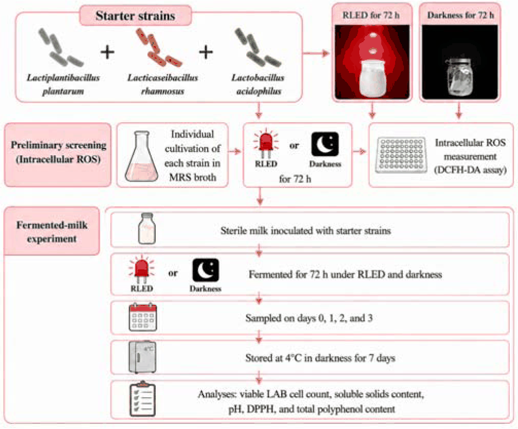
The RLED treatment was performed using a custom-designed irradiation system (ESLEDs, Seoul, Korea) equipped with a red LED light source with a peak wavelength of 660±5 nm. The distance between the LED light source and the sample surface was fixed at 10 cm throughout the experiment. Illuminance at the sample surface was measured using a multi-LED light meter (TM-209M, Tenmars Electronics, Taipei, Taiwan), and the spatial distribution of light was evaluated using LightTools software. The illuminance measured at the sample surface was 261.36 lux. Based on the CIE photopic luminous efficiency function for 660-nm light (CIE, 2019), the corresponding irradiance was estimated to be approximately 6.27 W/m² (0.627 mW/cm²). During fermentation, the RLED-treated samples were continuously exposed to red LED light for 72 h at 37℃, whereas the dark-control samples were incubated under the same temperature and environmental conditions without light exposure. Following 72 h of fermentation, RLED irradiation was discontinued, and the remaining samples from both treatment groups were stored at 4℃ in darkness. Samples were collected on days 1, 2, and 3 during fermentation and on days 6 and 10 during refrigerated storage for subsequent analyses.
Intracellular ROS-related responses were evaluated separately in Lactiplantibacillus plantarum, Lacticaseibacillus rhamnosus, and Lactobacillus acidophilus. Each strain was cultured in 6-well plates containing 5 mL of MRS broth at 37℃ for 72 h under RLED or dark conditions. After incubation, cells were collected by centrifugation at 10,000×g for 5 min at 4℃, washed twice with phosphate-buffered saline (PBS; pH 7.4), and resuspended in PBS. Cells were loaded with 2′,7′-dichlorodihydrofluorescein diacetate (DCFH-DA; D6883, Sigma- Aldrich, St. Louis, MO, USA) by mixing 1,350 µL of cell suspension with 150 µL of a 1 mM DCFH-DA stock solution prepared in dimethyl sulfoxide, yielding a final concentration of 100 µM. After incubation at 37℃ for 30 min in the dark, cells were washed twice with PBS and resuspended in PBS. For fluorescence measurement, 190 µL of the loaded cell suspension was transferred to a black 96-well plate. Hydrogen peroxide (H2O2) and N-acetyl-L-cysteine (NAC; A7250, Sigma-Aldrich, USA) were used as oxidative and antioxidant controls at final concentrations of 2 and 20 mM, respectively. PBS was used for the untreated assay condition and as the fluorescence blank. After incubation at 37℃ for 20 min, fluorescence was measured at 485 nm excitation and 535 nm emission using a microplate reader (SpectraMax iD5, Molecular Devices, San Jose, CA, USA). Fluorescence values were corrected by subtracting the PBS blank. Viable cell counts of the corresponding cultures were determined as described in the Viable cell count section.
Viable cell counts were determined using the standard plate count method. For preliminary evaluation of individual strain growth under RLED irradiation, activated cultures of Lactiplantibacillus plantarum, Lacticaseibacillus rhamnosus, and Lactobacillus acidophilus were separately inoculated into MRS broth at 2% (v/v) and cultivated at 37℃ under continuous RLED irradiation or dark conditions. Viable cell counts were determined at 0, 24, and 48 h. Fermented-milk samples collected during fermentation and subsequent refrigerated storage were analyzed using the same plate-count procedure. Individual LAB cultures and fermented-milk samples were serially diluted with sterile 0.85% (w/v) saline solution, and appropriate dilutions were spread onto MRS agar plates (Difco, BD, USA). The plates were incubated at 37℃ for 48–72 h, after which colonies were counted. Viable cell counts were expressed as log colony forming unit (CFU)/mL.
The soluble solids content (°Brix) of the fermented-milk samples was measured using a digital refractometer (PAL-S, ATAGO, Tokyo, Japan). Prior to measurement, the refractometer was calibrated with distilled water according to the manufacturer's instructions. Approximately 0.3 mL of each sample was applied to the prism surface, and the soluble solids content was recorded as °Brix. The prism surface was cleaned with distilled water and 70% (v/v) ethanol between measurements.
For pH determination, samples collected at each sampling time were stored at –80℃ until analysis and thawed at room temperature prior to measurement. The pH was measured using a digital pH meter (PAL-pH, ATAGO, Japan) after calibration with standard buffer solutions according to the manufacturer's instructions.
DPPH radical scavenging activity was determined according to the method of Blois (1958) with minor modifications. A 4 mM stock solution of 2,2-diphenyl-1-picrylhydrazyl (DPPH; Sigma-Aldrich, Taufkirchen, Germany) was prepared in ethanol and diluted to 400 µM immediately before use. For the assay, 100 µL of each sample was mixed with 100 µL of the DPPH working solution in a 96-well plate. The control consisted of 100 µL of DPPH solution mixed with 100 µL of ethanol (Daejung Chemicals & Metals, Siheung, Korea). The reaction mixtures were incubated at 37℃ for 10 min in the dark, and absorbance was measured at 517 nm using a microplate reader (SpectraMax iD5, Molecular Devices, USA). DPPH radical scavenging activity was calculated as follows:
where A_control and A_sample represent the absorbance values of the control and sample, respectively.
Total polyphenol content was determined using the Folin–Ciocalteu method according to Folin & Ciocalteu (1927) with minor modifications. The Folin–Ciocalteu reagent was diluted with distilled water at a ratio of 1:1 (v/v), and a 2% (w/v) Na2CO3 solution was prepared separately. For the assay, 80 µL of each sample was mixed with 20 µL of the diluted Folin–Ciocalteu reagent in a 96-well plate. After incubation for 3 min at room temperature, 100 µL of 2% (w/v) Na2CO3 solution was added, and the mixture was incubated at room temperature for 30 min. Absorbance was then measured at 750 nm using a microplate reader (SpectraMax iD5, Molecular Devices, USA). Tannic acid was used as the reference standard. A 1,000 µg/mL tannic acid stock solution was serially diluted two-fold to construct the standard curve. Total polyphenol content was calculated from the standard curve and expressed as tannic acid equivalents (TAE).
Data from the fermented-milk experiments and individual-strain DCFH-DA assays are presented as mean±standard deviation (SD) of three independent experimental replicates (n=3). Technical replicate measurements within each experimental replicate were averaged prior to statistical analysis. Differences among treatment–time combinations were analyzed by one-way analysis of variance (ANOVA), followed by Tukey's honestly significant difference (HSD) test for multiple comparisons. Because independent samples were collected at each treatment–time combination rather than by repeated measurement of the same samples over time, each combination was treated as an independent group and analyzed by one-way ANOVA. Comparisons of DCFH fluorescence between RLED and dark conditions for each individual LAB strain were performed using an independent Student's t-test. Statistical analyses were conducted using SPSS Statistics 24 (IBM SPSS, Armonk, NY, USA) and GraphPad Prism version 5.02 (GraphPad Software, San Diego, CA, USA). Differences were considered statistically significant at p<0.05. The individual-strain viable counts shown in Table 1 were obtained from a preliminary screening experiment intended to assess the growth responses of each strain to RLED prior to mixed-starter application and are presented as descriptive observations without statistical analysis.
Results and Discussion
Prior to applying the mixed starter culture to fermented milk, the three LAB strains were individually exposed to RLED to examine their responses to red-light treatment. As part of this preliminary screening, intracellular ROS-related changes were evaluated after 72 h of cultivation using the DCFH-DA assay (Fig. 2). DCFH fluorescence was significantly higher under RLED than under dark conditions in Lacticaseibacillus rhamnosus and Lactobacillus acidophilus (p<0.01), with the largest increase, approximately 26.5%, observed in L. acidophilus. In contrast, Lactiplantibacillus plantarum showed only a slight increase that was not statistically significant (p>0.05). The increase in fluorescence following H2O2 treatment and its marked decrease in the presence of NAC confirmed that the assay responded appropriately to changes in cellular oxidative status. Culturable counts of the three strains remained comparable between RLED and dark conditions, with differences of less than 0.1 log CFU/mL. Thus, the higher DCFH fluorescence observed in L. rhamnosus and L. acidophilus was associated with a change in intracellular redox status rather than an increase or loss of culturable cells. Visible light can interact with endogenous chromophores in microbial cells and alter cellular redox reactions. Porphyrins and flavins are among the major endogenous photosensitizers involved in light-induced ROS formation, although their photochemical activity is generally greater at shorter wavelengths, particularly in the blue region (Lipovsky et al., 2010; Plavskii et al., 2018). Responses to longer wavelengths, however, have also been reported. Polo et al. (1999) found that red-light exposure altered the physiology of Escherichia coli, including increases in growth rate and several enzyme activities, and proposed a role for singlet oxygen generated through excitation of intracellular chromophores. In the present screening experiment, the increase in DCFH fluorescence without a corresponding reduction in viable counts suggests that 660-nm irradiation acted as a sublethal cellular stimulus rather than causing photodynamic inactivation.
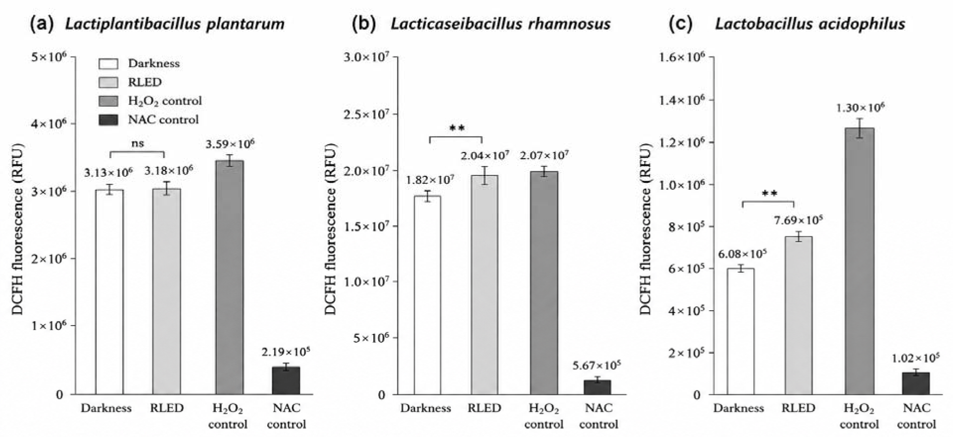
The extent of this response differed among the starter strains. LAB vary considerably in their ability to maintain redox homeostasis through NADH-dependent peroxide-removing systems, thiol-based pathways, and other antioxidant defenses (Kang et al., 2013; Bryukhanov et al., 2022). Strain-to-strain variation in antioxidant activity, glutathione content, superoxide dismutase activity, and resistance to oxidative stress has also been reported (Amaretti et al., 2013). Such physiological diversity may account for the clear ROS-associated responses of L. rhamnosus and L. acidophilus and the comparatively weak response of L. plantarum. These screening results indicate that the three starter strains did not respond uniformly to RLED and provided a basis for subsequently examining whether red-light exposure could influence the behavior of the mixed culture during fermented-milk production and refrigerated storage.
The individual starter strains were initially screened under RLED and dark conditions to determine their growth responses to red-light exposure before application as a mixed culture (Table 1). After 24 h of cultivation, Lacticaseibacillus rhamnosus reached 9.61 log CFU/mL under RLED compared with 9.19 log CFU/mL under darkness, while Lactobacillus acidophilus reached 9.14 and 8.37 log CFU/mL under RLED and dark conditions, respectively. Lactiplantibacillus plantarum showed similar viable counts under the two conditions at this time point. After 48 h, the difference between RLED and dark conditions was smaller for L. rhamnosus and L. acidophilus, whereas L. plantarum showed a lower viable count under RLED than under darkness. Together with the intracellular ROS-related results (Fig. 2), these observations show that the three starter strains exhibited different growth patterns under RLED and dark conditions over the cultivation period. Following this preliminary screening, the three strains were combined as a mixed starter and applied to fermented milk to examine whether RLED exposure could influence LAB viability during fermentation and subsequent refrigerated storage (Table 2). The initial population was 6.57 log CFU/mL. On day 1, the RLED-treated samples reached 8.02 log CFU/mL, whereas the dark control reached 7.51 log CFU/mL, indicating a more rapid early increase in the illuminated cultures. The pattern changed during prolonged fermentation, with higher viable counts in the dark control on days 2 and 3. After transfer to 4℃, the RLED-treated samples showed numerically higher viable counts, reaching 7.91 and 7.64 log CFU/mL on days 6 and 10, respectively, compared with 7.73 and 7.53 log CFU/mL in the corresponding dark controls, although these differences were not statistically significant at individual sampling times. Thus, the influence of RLED appeared to be dependent on the fermentation stage rather than being characterized by a continuous stimulation of cell growth.
Samples were fermented at 37℃ under dark or red light-emitting diode (RLED) conditions for 3 days and subsequently stored at 4℃ in darkness until day 10. Day 0 represents the baseline before initiation of RLED treatment. Values are presented as mean±SD (n=3). Different letters indicate significant differences among treatment–time combinations (p<0.05) according to Tukey's HSD test following one-way ANOVA.
Red-light photostimulation has also been reported to alter microbial populations in other fermentation systems. Martins et al. (2024) observed increased LAB and yeast populations during table olive fermentation under red LED illumination, suggesting that long-wavelength visible light can modify microbial growth behavior during fermentation. Although differences in microbial composition and fermentation matrices preclude direct comparison, these observations support the possibility that red light can influence the growth and physiological behavior of fermentative microorganisms. The strain-specific responses observed in the present screening further indicate that the outcome of RLED treatment is likely determined by the characteristics of the microorganisms comprising the starter culture. LAB can respond to moderate environmental stimuli by adjusting redox homeostasis and activating cellular stress-defense systems, and such physiological adaptation may influence responses to subsequent environmental changes (Papadimitriou et al., 2016). In the present study, RLED increased intracellular ROS-related fluorescence in L. rhamnosus and L. acidophilus without reducing their culturable populations. When the three strains were subsequently applied as a mixed starter culture, viable LAB counts showed different temporal patterns under RLED and dark conditions during fermentation and refrigerated storage. Although viable counts during refrigeration did not differ significantly between treatments at individual sampling times, the response to RLED varied with the stage of fermentation and storage. The relationship between the strain-specific redox response and the subsequent behavior of the mixed starter remains to be clarified; nevertheless, the present results show that 660-nm RLED was associated with strain-dependent cellular responses and changes in LAB growth patterns without a sustained loss of culturability. These findings suggest that RLED may serve as a non-thermal process variable for modulating starter behavior during fermented-milk production.
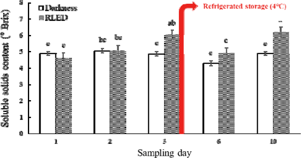
Changes in soluble solids content and pH were monitored to evaluate physicochemical changes during fermentation and subsequent refrigerated storage. The soluble solids content showed distinct temporal patterns between the two treatments (Fig. 3). In the dark control, the value increased slightly from 4.9 °Brix on day 1 to 5.1 °Brix on day 2, decreased to 4.3 °Brix on day 6, and returned to 4.9 °Brix by day 10. In contrast, the RLED-treated samples exhibited a greater range of variation, increasing from 4.6 °Brix on day 1 to 6.0 °Brix on day 3. Following refrigeration, the value decreased to 4.9 °Brix on day 6 and subsequently increased to 6.2 °Brix on day 10. These results indicate that RLED exposure was associated with a more pronounced temporal change in the soluble fraction of the fermented milk, particularly during the later stages of fermentation and refrigerated storage. Changes in soluble solids during lactic acid fermentation reflect the combined effects of substrate utilization and the formation of soluble fermentation products. LAB metabolize milk carbohydrates, primarily lactose, while producing organic acids and a variety of metabolites that alter the composition of the fermentation matrix. In addition, proteolytic activity and exopolysaccharide production by LAB may contribute to changes in soluble and colloidal components during fermentation (Cerning, 1990; Pham et al., 2000; Wang et al., 2021; Wu et al., 2023). Because these processes occur simultaneously, soluble solids do not necessarily change in a single direction over time. One possible explanation for the increase in soluble solids observed in the RLED-treated samples during refrigerated storage is the continuation of proteolytic activity by LAB-derived and residual enzymes at low temperature, which may gradually release low-molecular-weight peptides and free amino acids into the matrix (Serra et al., 2009; González-Olivares et al., 2014). The broader fluctuations observed under RLED may therefore reflect a different balance between substrate utilization and the formation or accumulation of soluble fermentation products; however, as °Brix represents a bulk measurement of total soluble constituents, these values do not directly identify the specific metabolic changes involved.
The pH decreased markedly in both treatment groups as fermentation progressed (Table 3). In the dark control, pH declined from 4.10 on day 1 to 3.58 on day 3 and remained within a narrow acidic range during refrigerated storage. The RLED-treated samples followed a comparable downward trend, decreasing from 4.25 on day 1 to 3.66 on day 3 and remaining near 3.57–3.60 through day 10; however, the pH of the RLED-treated samples was significantly higher than that of the dark control at each sampling point during fermentation (days 1–3, p<0.05), whereas both treatments remained within a narrow acidic range during refrigerated storage. This progressive acidification is consistent with the metabolic activity of LAB, which convert lactose into lactic acid and other organic acids during milk fermentation. The resulting decrease in pH contributes to casein destabilization and gel formation and is therefore an important determinant of the texture and physicochemical stability of fermented dairy products.
Samples were fermented at 37℃ under dark or red light-emitting diode (RLED) conditions for 3 days and subsequently stored at 4℃ in darkness until day 10. Values are presented as mean±SD (n=3). Different letters within each parameter indicate significant differences among treatment–time combinations (p<0.05) according to Tukey's HSD test following one-way ANOVA. TAE, tannic acid equivalents.
An important aspect of the present results is that the more pronounced changes in soluble solids under RLED were not accompanied by excessive acidification. Although significant differences in pH were observed between treatments during fermentation, both groups ultimately reached a similar acidic range and remained relatively stable during refrigerated storage. This suggests that RLED modulated the rate and course of acidification during fermentation while ultimately preserving the characteristic acidification pattern of LAB fermentation. When considered together with the viable-cell results, in which the RLED-treated samples tended to maintain higher LAB counts after transfer to 4℃, the data indicate that red-light exposure influenced starter performance and fermentation behavior without disrupting the overall acid–base balance of the product. The combined behavior of soluble solids and pH therefore provides complementary evidence that RLED affected the progression of fermentation rather than simply accelerating or suppressing microbial activity. The greater variability in °Brix under RLED may be consistent with altered substrate conversion and metabolite formation, whereas the comparable final pH range indicates that the fundamental acidification process remained well controlled. It should be noted, however, that °Brix reflects the total soluble solids content and does not by itself demonstrate specific metabolic transformations. This combination may be advantageous from a processing perspective, as RLED was associated with changes in fermentation-related biochemical activity and a tendency toward better maintenance of LAB viability during refrigeration without causing marked post-fermentation acidification.
DPPH radical scavenging activity changed during fermentation and subsequent refrigerated storage in both treatment groups (Fig. 4). In the dark control, activity increased from 65.1% on day 1 to 85.1% on day 3 and then declined during refrigerated storage. The RLED-treated samples showed a different temporal pattern, increasing from 64.6% on day 1 to 81.2% on day 3 and maintaining a similar level on day 6 (81.6%) before decreasing to 70.5% on day 10. Thus, antioxidant activity increased during the fermentation period in both groups, whereas the subsequent pattern differed after transfer to refrigeration. In particular, the RLED-treated samples maintained relatively high radical scavenging activity through the early stage of refrigerated storage.
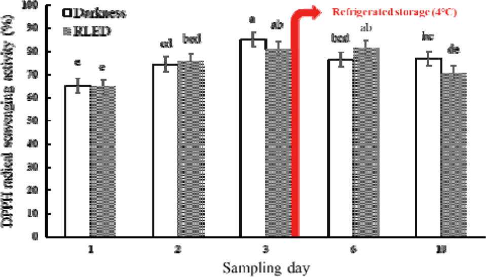
The increase in antioxidant activity during milk fermentation can arise from several biochemical processes associated with LAB metabolism. Proteolysis of milk proteins may release peptides and amino acids with radical-scavenging properties, while milk proteins and sulfur-containing amino acids can also contribute to the antioxidant capacity of dairy products (Corrochano et al., 2018). Microbial metabolism may further alter the composition and redox properties of low-molecular-weight compounds present in the milk matrix. Accordingly, the increase in DPPH activity observed during fermentation is consistent with the progressive formation and transformation of antioxidant-active components. The persistence of high activity in the RLED-treated samples through day 6 suggests that red-light exposure influenced the temporal development of these fermentation-related antioxidant properties.
Total polyphenol content decreased markedly from the day-0 baseline of 28.83 µg TAE/100 g to day 1 in both treatments (Table 3). Thereafter, the values remained within a considerably narrower range. In the dark control, total polyphenol content ranged from 11.10 to 11.88 µg TAE/100 g, whereas the RLED-treated samples increased from 10.22 µg TAE/100 g on day 1 to 12.26 µg TAE/100 g on day 6 before decreasing slightly to 11.62 µg TAE/100 g on day 10. The increase observed in the RLED-treated samples through day 6 occurred over a similar period in which relatively high DPPH radical scavenging activity was observed.
Lactic acid fermentation can modify phenolic-related compounds through microbial enzymatic reactions. LAB are capable of hydrolyzing conjugated compounds and converting glycosylated forms into aglycones or other derivatives with altered chemical properties (Chien et al., 2006; Park et al., 2019). Changes in phenolic composition during lactic fermentation have also been associated with enhanced antioxidant characteristics in various fermented food systems (Ryu et al., 2018; Song et al., 2020; Lim, 2022). In dairy matrices, interactions between phenolic compounds, milk proteins, and microbial metabolites may further influence the measurable polyphenol content during fermentation and storage. The increase observed in the RLED-treated samples up to day 6 therefore suggests that red-light exposure may have affected the transformation or availability of phenolic-related components during the course of fermentation, although the Folin–Ciocalteu assay reflects total reducing capacity rather than specific phenolic transformations and therefore does not directly identify the compounds involved. The temporal profiles of DPPH scavenging activity and total polyphenol content were not completely parallel. This difference is reasonable because antioxidant activity in fermented milk is determined by multiple constituents rather than phenolic compounds alone. Milk-derived peptides, amino acids, thiol-containing compounds, endogenous antioxidants, and microbial metabolites can all participate in radical-scavenging reactions. The two measurements therefore provide complementary rather than interchangeable information on the functional changes occurring in the fermented matrix.
The antioxidant-related indices therefore indicate that RLED exposure influenced the temporal development of functional properties during fermentation and refrigerated storage. The RLED-treated samples maintained high DPPH radical scavenging activity into the early refrigerated-storage period, while total polyphenol content increased up to day 6 before declining slightly thereafter. When considered together with the changes in LAB viability, these findings suggest that the influence of RLED extended beyond microbial growth and was accompanied by changes in the functional characteristics of the fermented milk. This response supports the potential application of RLED as a non-thermal processing approach for modulating both starter performance and antioxidant-related quality during fermented dairy production.
Conclusion
This study examined the response of three LAB starter strains to 660-nm RLED irradiation and subsequently evaluated the effects of RLED treatment on fermented milk during fermentation and refrigerated storage. Preliminary screening revealed strain-dependent responses to red light. In particular, RLED increased intracellular ROS-related fluorescence in Lacticaseibacillus rhamnosus and Lactobacillus acidophilus without reducing their culturable populations, indicating that red-light exposure can alter cellular redox status without compromising viability under the conditions tested. When the three strains were combined as a starter culture, RLED treatment affected the temporal pattern of LAB growth and was associated with numerically higher viable counts during subsequent refrigerated storage, although these differences were not statistically significant. This raises the possibility that the physiological responses induced during light exposure persist beyond the irradiation period, which remains to be confirmed. RLED was also associated with changes in soluble solids while preserving the characteristic acidification profile of fermented milk, indicating that its effects on microbial activity were not accompanied by excessive post-fermentation acidification. Changes in DPPH radical scavenging activity and total polyphenol content further indicated that the influence of RLED extended to antioxidant-related characteristics of the fermented matrix, with antioxidant activity maintained into the early refrigerated-storage period. The present findings therefore position RLED as a potential process variable for modulating LAB physiology and fermentation behavior rather than simply as a means of stimulating microbial growth. Its apparent ability to influence starter performance while maintaining key physicochemical characteristics provides a basis for the development of light-assisted fermentation processes in dairy systems. Further studies are needed to optimize irradiation conditions, including wavelength, light intensity, exposure time, and starter composition, and to confirm the reproducibility of the observed responses under practically relevant fermentation conditions. These findings provide a basis for further evaluating RLED as a non-thermal process variable for modulating starter behavior and fermentation characteristics in fermented dairy systems.


