Reaserch article

식품착색제 이산화티타늄 유발 DNA 손상에 대한 쓴메밀 추출물의 in vitro 항산화 효과

송윤서1https://orcid.org/0009-0006-2143-0888, 최수진1,*https://orcid.org/0000-0001-5131-8156
Yoon-Seo Song1https://orcid.org/0009-0006-2143-0888, Soo-Jin Choi1,*https://orcid.org/0000-0001-5131-8156
Author Information & Copyright
1서울여자대학교 식품생명공학과
1Department of Food Science and Biotechnology, Seoul Women’s University, Seoul 01797, Korea
*Corresponding author: Soo-Jin Choi, Department of Food Science and Biotechnology, Seoul Women’s University, Seoul 01797, Korea, E-mail: sjchoi@swu.ac.kr

ⓒ Copyright 2026 Korean Society for Food Engineering. This is an Open-Access article distributed under the terms of the Creative Commons Attribution Non-Commercial License (http://creativecommons.org/licenses/by-nc/4.0/) which permits unrestricted non-commercial use, distribution, and reproduction in any medium, provided the original work is properly cited.

Received: Jul 23, 2026; Revised: Aug 06, 2026; Accepted: Aug 03, 2026

Published Online: Sep 03, 2026

Abatract

Tartary buckwheat (Fagopyrum tataricum), named for its relatively bitter taste compared with common buckwheat, has attracted attention as a functional food due to its superior nutritional value, characterized by substantially higher levels of rutin, total polyphenols, and total flavonoids. Titanium dioxide (TiO2), widely used as a food colorant, remains controversial because of its potential genotoxicity. When used as a food additive, its toxicological impact may vary depending on interactions with food matrices or other nutritional components. This study evaluated TiO2-induced oxidative stress and DNA damage in intestinal cells. Furthermore, we investigated the inhibitory effects of Tartary buckwheat extract (TBE) and its major bioactive compound rutin against the oxidative stress-induced genotoxicity. The results demonstrated that TiO2 generated reactive oxygen species and induced oxidative stress that led to DNA damage. Notably, TBE, which possesses potent antioxidant properties such as rutin, mitigated the oxidative stress and subsequent DNA damage. These findings provide foundational data on the potential genotoxicity of TiO2 as a food colorant and on the protective effects of antioxidant compounds against genotoxicity.

Keywords: Tartary buckwheat; Antioxidant effect; Titanium dioxide; Oxidative stress; DNA damage

서 론

메밀(Fagopyrum esculentum)은 마디풀(Polygonaceae)과에 속하는 관속식물로 그 종실은 농경지에서 식용 곡물로 재배될 뿐만 아니라 산림 주변부 및 제방 등에 야생화하여 자라는 피복작물(cover crop)로도 이용된다. 중국 남중부와 티베트가 원산지이고, 생육기간이 60~80일로 서늘하고 습한 기후에서 재배되며, 척박한 토양 조건에서도 잘 자라 동유럽, 중앙아시아, 동아시아, 북미 등 전 세계에서 널리 재배되고 있다(Worthing et al., 2025). 동유럽과 중앙아시아에서는 알곡을 그대로 익혀 먹거나 죽 형태로, 서유럽과 북미에서는 가루를 낸 뒤 반죽하여 부쳐 먹는 팬케이크나 크레페 형태로, 동아시아와 이탈리아 북부 지역에서는 냉면, 막국수, 소바, 파스타 등의 면 형태로 소비되고 있다(Huda et al., 2021; Ranic et al., 2025). 메밀에는 루틴(rutin, quercetin-3-o-rutinoside), 퀘르세틴(quercetin) 등 항산화 성분과 D-chiro-inositol과 같이 인슐린 신호 전달을 강화해 인슐린 저항성을 개선하고 혈당을 조절하는 생리활성 성분 및 양질의 단백질, 비타민 B군, 미네랄, 식이섬유 등 다양한 영양성분을 함유하고 있다(Zhu, 2016; Huda et al., 2021). 따라서 메밀은 혈압 조절, 뇌졸중 예방, 당뇨 조절, 간 해독 및 기능 활성, 장 건강 개선, 다이어트, 항산화 및 항염증 등의 목적으로 널리 섭취되어 왔다(Nakamura et al., 2013; Zhu, 2016; Huda et al., 2021).

쓴 메밀(Fagopyrum tataricum)은 일반 메밀보다 줄기가 가늘고 꽃의 크기가 작고, 주로 히말라야 산맥, 중국 사천성 등 고산지대에서 척박한 환경을 견디며 자라는 특성이 있으며, Fagopyrum esculentum과 같은 마디풀과에 속한다. 일반 메밀보다 쓴맛이 강해 ‘쓴 메밀’이라 불리고, 메밀의 핵심 기능성 성분인 rutin 함량이 일반 메밀보다 약 40~100배 이상 높은 것으로 알려져 있다(Aubert et al., 2021; Song et al., 2022). 또한 총 폴리페놀 및 총 플라보노이드 함량이 일반 메밀보다 월등히 높은 것으로 보고되고 있어 탁월한 영양학적 가치와 생리활성 덕분에 ‘슈퍼 푸드’로 각광받고 있다(Mazahir et al., 2022; Jha et al., 2024).

식품첨가물인 이산화티타늄(TiO2)은 티타늄과 산소가 반응하여 형성되는 산화물로, 식품 산업에서 백색도와 광택을 높이는 착색제(coloring agent)로서 사탕, 껌, 제과류, 커피 크리머, 초콜릿, 유제품, 소스 및 드레싱 등에 사용되고 있다. TiO2는 자연적으로 발생하는 무기 불용성 안료로서 물리적, 화학적 및 생물학적 방법을 통해 합성될 수 있다(Irshad et al., 2021). TiO2는 1966년 미국식품의약국(United States Food and Drug Administration, FDA)의 21 CFR 73.575에 따라 식품에서 사용이 처음으로 허가되었으며, 식품의 중량 중 1%를 초과하지 않도록 권고되고 있다(FDA, 2024). 2008년에는 유럽연합(European Union, EU) 규정(European Commission, EC) No. 1333/2008에 따라 식품첨가물 E171로 승인된 바 있다(EUR-Lex, 2008). 그러나, 2021년 유럽 식품안전청(European Food Safety Authority, EUR-Lex)은 E171에 대한 안전성을 재평가하여, 잠재적인 유전독성 가능성을 배제할 수 없으므로 2022년 식품첨가물로서의 사용을 금지하였다(EFSA, 2022). 한편, in vivo 생체 수준에서의 TiO2의 유전독성에 대한 과학적 근거자료가 명확하지 않아 미국 FDA, 캐나다 보건부(Health Canada), 영국 식품기준청(Food Standards Agency), 호주·뉴질랜드 식품기준청(Food Standards Australia New Zealand) 및 우리나라에서는 식품첨가물로서 안전성에 문제가 없는 것으로 판단하고 있다(FSA, 2022; FSANZ, 2022; Health Canada, 2022; MSDS, 2025). 특히, 식품첨가물로서 사용 시 식품 원료나 다른 영양성분들과의 상호작용에 의해 독성영향이 달라질 수 있다(Jo et al., 2016; Jeong et al., 2025). 착색제 TiO2의 산화스트레스 유발과 in vitro 유전독성에 대한 연구 및 쓴메밀의 기능성에 대한 결과는 각각 보고된 바 있으나, 쓴메밀 섭취에 의한 TiO2 유전독성 완화 효과에 대해서는 보고된 바 없다.

따라서, 본 연구에서는 식품첨가물 TiO2의 물리화학적 특성 분석을 기반으로 장관세포주에서 산화스트레스 유발에 의한 DNA 손상 정도를 규명하고, 쓴메밀 추출물(Tartary buckwheat extract, TBE)과 이의 주된 기능성 성분인 rutin에 의한 TiO2 유발 산화스트레스 및 유전독성 완화 영향을 연구하고자 하였다.

재료 및 방법

메밀 및 추출물 제조

본 연구에 사용된 쓴메밀은 (주)봉평농원 (Pyeongchang, Korea)에서 구입하였다. 쓴메밀 10 g에 70% 에탄올(Samchun pure chemical Co., Ltd., Pyeongtaek, Korea) 200 mL를 가한 뒤 40℃에서 30분간 초음파 추출(Bransonic 5800, Branson Ultrasonics, Danbury, CT, USA)하여, 4℃에서 원심분리(14,000×g, 20분) 후 얻은 상등액을 TBE로 사용하였다(Yu et al., 2018; Jin et al., 2020).

식품첨가물 TiO2 특성 분석

두 가지 다른 상업용 식품첨가물 TiO2 (T3, T4)는 제조사로부터 제공받아 사용하였으며, 선행문헌의 결과에 따라 T3와 T4를 대표적 입자크기가 큰 물질과 작은 물질로 각각 선정하였다(Hwang et al., 2019). TiO2 입자는 실험 직전 1 mg/mL의 농도로 distilled deionized water (DDW)에 30분간 stirring하여 분산 후, 5분 동안 초음파 처리(160 Watts, Bransonic 5800, Branson Ultrasonics)하여 사용하였다.

TiO2의 구성 입자 크기 및 모양은 전계방출형 주사전자현미경(field emission-scanning electron microscopy, FE-SEM; JSM-7100F, JEOL, Tokyo, Japan)으로 분석하였다. 카본 테이프(5 mm×5 mm, E-SONG EMC, Seoul, Korea)가 부착된 마운트(Specimen Mount, JEOL) 위에 TiO2 용액(20 mL, 0.1 mg/mL)을 올리고 실온에서 건조한 후, 샘플 표면을 30초 동안 Pt/Pd로 코팅하여 15 kV의 가속 전압에서 FE-SEM 이미지를 획득하였다. TiO2 현탁액(0.1 mg/mL)의 유체역학적 직경(hydrodynamic diameters)과 제타 전위(zeta potentials)는 Zetasizer Nano 시스템(Malvern Instruments, Worcestershire, UK)을 이용하여 각각 동적 광산란법(dynamic light scattering)과 전기영동 광산란법(electrophoretic light scattering)으로 DDW와 세포 배양액 minimum essential medium (MEM)에서 각각 측정하였다. TBE와 rutin (Sigma-Aldrich, St. Louis, MO, USA) 존재하 TiO2 입자의 유체역학적 직경은, rutin 농도가 14.2 mM이 되도록 첨가하여 측정하였는데, 이는 선행결과 rutin이 세포독성을 나타내지 않는 농도이다(Jin et al., 2020).

Rutin 및 quercetin 함량 측정

TBE 내 rutin 및 quercetin (Sigma-Aldrich)의 함량은 주사기 필터(0.45 µm; Advantec®, Toyo Roshi Kaicha Ltd., Tokyo, Japan)로 여과한 뒤, high performance liquid chromatography (HPLC)로 diode-array detector를 이용하여 분석하였다. 고정상 SupelcosilTM LC-18 (250 mm×4.6 mm i.d., 5 µm, Supelco Inc., Bellefonte, PA, USA) 컬럼을 사용하여 1 mL/min 유속으로 20 µL를 주입하였고, methanol (Daejung chemical & metals Co., Ltd., Siheung, Korea), acetonitrile (Samchun pure chemical Co., Ltd.), 2.5% acetic acid (JT Baker, Phillipsburg, NJ, USA)를 1:2:7 (v/v/v) 비율로 혼합한 이동상으로 350 nm 파장에서 흡광도를 측정하여 정량분석하였다.

총 폴리페놀 및 총 플라보노이드 함량 측정

TBE의 총 폴리페놀 함량은 Folin-Denis법(Appel et al., 2001)에 따라 분석하였다. TBE (80 µL, 156 µg/mL)에 50% Folin-Ciocalteu 시약(20 µL, Sigma-Aldrich)을 가하여 암소에서 5분간 반응시킨 후, 2% Na2CO3 용액(100 µL, Samchun Pure Chemical Co., Ltd.)을 가하여 30분 동안 암소에서 처리하여 microplate reader (SpectraMax® M3, Molecular Devices, Sunnyvale, CA, USA)로 750 nm에서 흡광도를 측정하였다. 총 폴리페놀의 함량은 tannic acid (Sigma-Aldrich)을 표준물질로 이용한 표준곡선으로부터 계산하였다.

TBE의 총 플라보노이드 함량은 Zhishen et al.의 변형된 방법에 따라 측정하였는데(Zhishen et al., 1999), TBE (50 µL)에 5% NaNO2 용액(30 µL, Sigma-Aldrich)을 가하여 5분간 반응시킨 후, 2% AlCl3·6H2O 용액(60 µL, Sigma-Aldrich)를 첨가하여 암소에서 6분 동안 반응시켰다. 1 N NaOH 용액(100 µL, Duksan Pure Chemicals Co., Ltd., Ansan, Korea)을 첨가하여 11분간 반응시킨 뒤, 510 nm에서 흡광도를 측정하였다(SpectraMax® M3, Molecular Devices). 총 플라보노이드의 함량은 (+)-catechin (ENZO Life Sciences, Farmingdale, NY, USA)을 표준물질로 이용한 표준곡선에 의해 계산하였다. 상기의 두 실험 모두 TBE 내 rutin 함량을 비교군으로 이용하였다.

항산화 활성 측정

TBE의 항산화 활성은 2,2'-azinobis-(3-ethyl-benzothiazoline-6-sulfonic acid) (ABTS; Sigma-Aldrich)를 이용하여 측정하였는데(Lee et al., 2013; Lee et al., 2015), 10 mM ABTS 용액과 10 mM K2S2O8 용액을 7.4:2.6 (v/v)의 비율로 혼합한 후, 37℃ 암소에서 24시간 반응시켜 ABTS 라디칼을 형성하고, 인산완충용액(phosphate buffered saline, PBS)으로 734 nm에서 0.65±0.02의 흡광도가 되도록 희석하였다. TBE 50 µL에 희석한 ABTS 라디칼 용액 150 µL를 가하여 상온 암소에서 30분간 반응시키고 734 nm에서 흡광도를 측정하였다(SpectraMax® M3, Molecular Devices).

2,2-diphenyl-2-picrylhydrazyl (DPPH; Alfa Aesar, Wardhill, MA, USA) 라디칼 소거능 분석에 의한 TBE의 항산화 활성 또한 측정하였다(Lee et al., 2013). 즉, 600 µM의 DPPH 용액을 메탄올에 제조한 뒤, 80% (v/v) 메탄올을 이용하여 517 nm에서 0.65±0.02의 흡광도가 되도록 희석하였다. TBE 100 µL와 희석한 DPPH 용액 100 µL를 혼합하여 암소에서 30분간 반응시킨 후 517 nm에서 흡광도를 측정하였다(SpectraMax® M3, Molecular Devices). TBE의 항산화 활성은 L-ascorbic acid (Samchun Pure Chemical Co., Ltd.)에 해당하는 항산화력으로 나타내었다. 상기의 두 실험 모두 TBE 내 rutin 함량을 비교군으로 이용하였다.

세포배양

Caco-2 장관상피 세포주를 한국세포주은행(Korean Cell Line Bank, Seoul, Korea)에서 분양 받아 10% 열 불활성화 fetal bovine serum, 페니실린(100 units/mL) 및 스트렙토마이신(100 µg/mL)을 함유한 MEM (Welgene Inc., Gyeongsan, Korea)에 5% CO2, 37℃ 조건의 세포배양기에서 배양하였다.

세포독성

Caco-2 세포에서의 성장 저해 독성은 water-soluble tetrazolium salt-1 (WST-1; Roche, Basel, Switzerland) 분석법으로 평가하였다. Caco-2 세포(1×10⁴ 세포/100 µL)에 TiO2를 단독 또는 TBE와 함께 24시간 처리하였는데, 최고 농도 292 µg/mL는 선행연구결과에 기반하여 다음과 같이 설정하였다: 품목제조보고서 기반 최대 사용량×일일섭취량 (g)/장관 부피 (100 mL) (Mudie et al., 2014; Kim et al., 2021). TBE 농도는 선행결과 세포독성이 나타나지 않는 농도로 설정하였는데, rutin 함량 기준 14.2 mM에 해당하며, 같은 농도의 표준물질 rutin을 비교 대조군으로 이용하였다(Jin et al., 2020). WST-1 시약(10 µL)을 첨가하여 4시간 동안 배양한 후, microplate reader (Infinite® M Plex, Tecan, Männedorf, Switzerland)를 이용하여 440 nm에서 흡광도를 측정하였다. 세포독성은 3반복 후 재현성을 다시 확인(n=3)하여 평균±표준편차로 나타내었다.

세포 내 활성산소종 생성 및 항산화효소 활성

세포 내 생성된 활성산소종(reactive oxygen species; ROS)은 세포막 투과성 2',7'-dichlorofluoroescein diacetate (H2DCFDA; Molecular Probes Inc., Eugene, OR, USA)를 이용하여 분석하였다. Caco-2 세포(1×10⁴ 세포/100 µL)에 TiO2를 단독으로 6시간 처리하거나, TBE 또는 rutin을 18시간 선처리하고 TBE 또는 rutin 존재하에 TiO2를 6시간 처리한 후, 비형광성 H2DCFDA (20 µM, 100 µL)를 첨가하여 암소에서 30분간 반응시켰다. PBS로 세척한 후, 세포 내 esterase에 의해 탈아세틸화되고 ROS와 반응하여 형광을 나타내는 dichlorofluorescein (DCF)를 excitation 파장과 emission 파장 각각 485 nm와 535 nm에서 측정하였다(Infinite® M Plex, Tecan).

산화스트레스의 지표로서 catalase (CAT)와 superoxide dismutase (SOD) 활성은 세포(1×10⁶ 세포/2 mL)에 TiO2를 단독으로 6시간 처리하거나, TBE 또는 rutin을 18시간 선처리하고 TBE 또는 rutin과 함께 TiO2를 6시간 처리한 후, 각각 CAT와 SOD 분석 kit (Cayman Chemical Co., Ann Arbor, MI, USA)를 이용하여 제조사의 프로토콜에 따라 분석하였다. 모든 실험은 3반복 후 재현성을 다시 확인(n=3)하여 평균±표준편차로 나타내었다.

In vitro 코멧 분석(comet assay)

Caco-2 세포(1×10⁶ 세포/2 mL)에 TiO2를 단독 처리하거나, TBE 또는 rutin을 18시간 선처리한 후 TBE 또는 rutin 존재 하에 TiO2를 6시간 추가 노출시켰고, 양성대조군으로 H2O2 (100 µM)를 이용하였다. Ethylene-diamine-tetraacetic acid 용액 (5 mM, 1 mL)을 40초 동안 처리하여 스크래퍼로 회수한 세포를 원심 분리하고 차가운 PBS 용액에 현탁(1×10⁵ 세포/1 mL)한 후, R&D Systems (Minneapolis, MN, USA)의 CometAssay® kit를 이용하여 제조사의 프로토콜에 따라 분석하였다. 최종적으로 Axioskop 2 plus 형광현미경과 Cometscore 2.0 이미지 분석 소프트웨어(Carl Zeiss, Oberkochen, Germany)를 이용하여 Comet 슬라이드에 대한 디지털 이미지 분석을 통해 comet tail의 길이를 측정하였으며, 전체 DNA 중 tail이 차지하는 DNA 백분율(%)을 수식[(tail 내 DNA 양/총 DNA 양)×100]에 의거하여 도출하였다. Comet 분석은 3반복 후 재현성을 다시 확인(n=3)하여 평균±표준편차로 나타내었다.

통계분석

모든 실험 데이터는 평균값±표준편차로 표기하였으며, Statistical Analysis System 소프트웨어(Version 9.4, SAS Institute, Inc., Cary, NC, USA)를 이용하여 일원배치 분산분석(one-way ANOVA)을 실시하였다. 각 군 간의 유의성 검정은 Tukey의 다중비교 검증을 통해 p<0.05 수준에서 실시하였다.

결과 및 고찰

TiO2의 물리화학적 특성

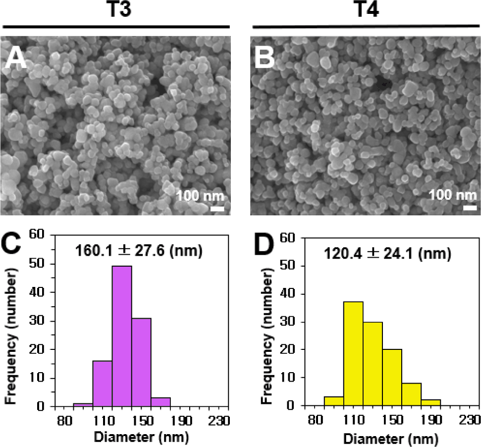
두 가지 식품첨가물 TiO2의 구성입자 및 모양을 FE-SEM을 이용하여 분석한 결과, 구형의 불규칙한 모양으로 관찰되었고, 평균 구성입자크기는 T3는 160.1±27.6 nm, T4는 120.3±24.1 nm로 측정되었다(Fig. 1, Table 1). 특히, T3의 경우 100 nm 미만의 입자가 관찰되지 않고 100~200 nm 크기가 99%를 구성하고 있는 것으로 나타난 반면, T4는 100 nm의 입자가 17% 존재하여 입자분포도에 있어서 차이가 있는 것으로 측정되었다. 즉, T3가 T4에 비하여 구성입자 크기가 유의적으로 더 큰 것을 확인할 수 있었다(Hwang et al., 2019; Jeong et al., 2025). 나노물질 정의에 따르면 평균 입자크기의 50% 이상이 1~100 nm 범위에 해당해야 나노물질로 분류된다(EFSA, 2016). 이러한 기준에 비추어 볼 때, 본 결과는 식품첨가물 TiO2가 나노물질에 해당되지 않음을 의미하며, 이는 선행문헌과 일치하는 결과이다(Hwang et al., 2019; Jeong et al., 2025).
fep-30-0-260034-g1
Fig. 1. (A, B) Field emission-scanning electron microscopic (FE-SEM) images and (C, D) constituent particle size distributions of food-grade TiO2 particles (T3 and T4). Particle size distribution was analyzed by measuring 100 randomly selected particles from the FE-SEM micrographs.
Download Original Figure
Table 1. Average particle size and size distribution of food-grade TiO2 particles (T3, T4) measured by FE-SEM
Sample Average size(nm) Distribution (Number %)
100 nm 100-200 nm >200 nm
T3 160.1±27.6a ND 99 1
T4 120.3±24.1b 17 83 ND

Different lower-case letters (a,b) next to the values indicate significant difference between TiO2 particles (T3, T4) (p<0.05). ND, not detected.

Download Excel Table

두 물질의 유체역학적 직경은 DDW와 세포배양 조건을 감안한 MEM에서 각각 측정하였으며, 두 물질 모두 DDW 분산 시보다 MEM 분산의 경우 유체역학적 직경이 유의적으로 증가하는 것으로 나타났다(Table 2, p<0.05). 이는 세포배양액 내 단백질, 아미노산 및 무기물질 등 다양한 영양성분과 TiO2가 상호작용하여 입자표면에 코로나(corona)를 형성했기 때문인 것으로 판단된다(Peng et al., 2015; Gräfe et al., 2016; Jeong et al., 2025). 한편, TiO2의 유체역학적 직경은 DDW와 MEM 조건에서 모두 TBE 존재 시 유의적으로 증가하였으나, rutin이 존재할 경우 유의적 감소하는 것으로 나타났다(p<0.05). 이는 rutin과 상호작용을 통해 TiO2의 분산 안정성이 향상될 수 있음을 의미하며, rutin이 capping agent로 작용하여 무기 나노입자를 안정화한다는 선행 연구결과와 일치한다(Joo & Chung, 2019; Łabuz et al., 2019). 한편, TiO2의 유체역학적 직경이 FE-SEM에 의한 구성입자크기 대비 크게 증가하여 응집된 상태로 존재하는 것으로 나타났으며, 이는 실질적으로 식품이나 생체 내에서는 벌크한 응집체로 존재함을 의미한다.

Table 2. Hydrodynamic diameters and zeta potentials of TiO2 particles (T3, T4) dispersed with TBE or rutin trihydrate in DDW or MEM
Sample Z-average size (nm) Zeta potential (mV)
T3 DDW T3 only 303.3±4.1Aa -35.8±0.2a
T3+TBE 316.2±0.5Ac -37.8±0.9c
T3+rutin 280.1±2.4Ab -30.4±0.6c
MEM T3 only 454.6±2.5Ba -8.7±0.4c
T3+TBE 490.9±3.0Bc -10.4±1.3a
T3+rutin 407.0±5.1Bb -8.8±1.9b
T4 DDW T4 only 293.0±5.2Aa -37.4±0.7a
T4+TBE 310.8±0.9Ac -33.7±0.1c
T4+rutin 276.6±3.0Ab -30.7±0.8b
MEM T4 only 412.2±2.4Ba -9.7±1.3a
T4+TBE 429.7±1.7Bc -10.3±1.7a
T4+rutin 372.4±7.8Bb -6.8±0.5b

Different uppercase letters (A,B) next to the values denote significant differences in hydrodynamic diameters between TiO2 dispersed in DDW and that in MEM (p<0.05). Different lowercase letters (a~c) next to the values indicate significant differences among TiO2 only, TiO2 with TBE, TiO2 with rutin prepared in either DDW or MEM (p<0.05). TBE, Tartary buckwheat extract; DDW, distilled and deionized water; MEM, minimum essential medium.

Download Excel Table
TBE의 기능성 성분 및 항산화활성 분석

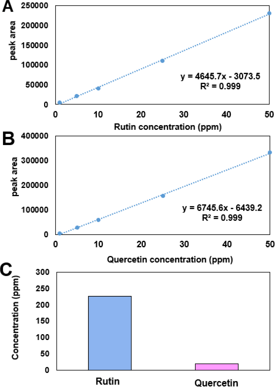
HPLC를 이용하여 TBE의 주요 기능성 성분인 rutin 함량과, 메밀 씨앗 내 rutin 3-glucosidase의 작용으로 rutin이 분해되어 생성되는 quercetin의 함량을 분석하였다. 그 결과, rutin과 quercetin 함량은 각각 229 ppm과 19 ppm으로 분석되었고(Fig. 2), rutin이 주된 기능성 성분으로 존재하고 있음을 확인하였다(Jin et al., 2020).
fep-30-0-260034-g2
Fig. 2. Standard curves for (A) rutin and (B) quercetin, and (C) the quantification of these compounds in TBE by HPLC.
Download Original Figure

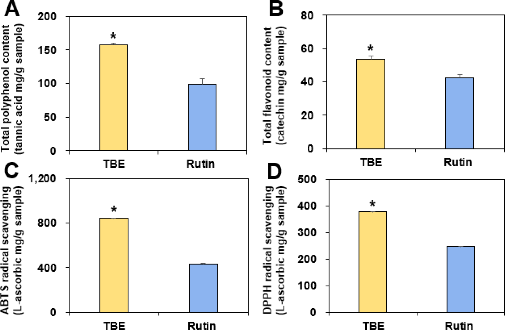
또한 TBE는 항산화 성분을 다량 함유하고 있는 것으로 알려져 있어 총 폴리페놀 및 총 플라보노이드 함량을 분석하였다. 이때, rutin이 폴리페놀계 플라보노이드 화합물이므로, HPLC로 정량한 TBE 내 rutin 함량에 해당하는 동량의 rutin을 비교군으로 사용하였다. 그 결과, TBE의 총 폴리페놀 및 총 플라보노이드 함량이 rutin 대비 약 1.6배 및 1.3배 높은 것으로 나타나(Fig. 3A, B), TBE 내 rutin 외 다른 폴리페놀 및 플라보노이드 성분들이 존재함을 알 수 있다(Jin et al., 2020; Vieira et al., 2022). 향후 이러한 다양한 폴리페놀 및 플라보노이드 성분에 대한 LC-MS 분석 기반의 프로파일링 연구가 수행되어야 할 것으로 보인다.
fep-30-0-260034-g3
Fig. 3. (A) Total polyphenol and (B) total flavonoid contents, and (C) ABTS and (D) DPPH radical scavenging activities of TBE compared with an equivalent amount of rutin standard. *Denotes significant differences between TBE and the rutin standard (p<0.05).
Download Original Figure

항산화 활성으로 ABTS 및 DPPH 라디칼 소거능을 분석한 결과, TBE의 라디칼 소거능이 rutin 대비 각각 약 2.0배 및 1.5배 높은 것으로 나타났는데(Fig. 3C, D), 이 역시 rutin 외 다른 기능성 성분들이 ABTS 및 DPPH 라디칼 소거능에 기여하고 있음을 보여준다.

TBE의 TiO2 유도 ROS 생성 저해 및 항산화효소 활성 증진 효과

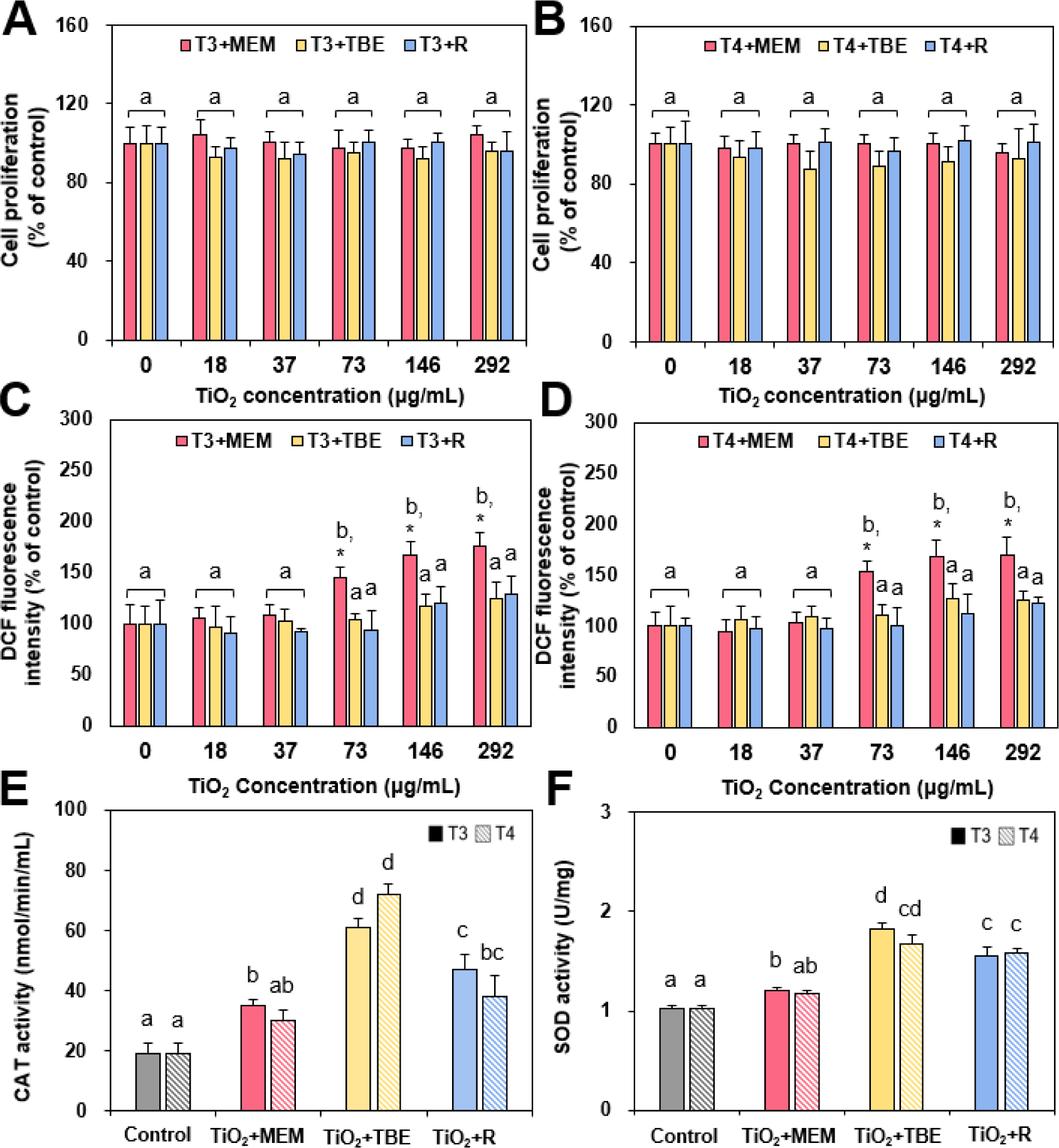
선행연구에서 식품첨가물 TiO2는 사용량 기반 최고 농도인 292 µg/mL까지 세포성장을 저해하거나 사멸을 유발하지 않는 것으로 보고되었으나(Jeong et al., 2025), 본 연구에서는 TiO2와 TBE 또는 rutin과의 상호작용이 세포독성에 미치는 영향을 세포성장 저해 관점에서 추가 평가하였다. 그 결과, 두 가지 TiO2 단독 처리 시(세포배양액 MEM 조건)와 TBE 또는 rutin과 함께 24시간 처리할 경우 모두 세포성장 저해를 유발하지 않았다(Fig. 4A, B).
fep-30-0-260034-g4
Fig. 4. (A, B) Effect of the presence of TBE or rutin on cell proliferation inhibition caused by TiO2 particles (T3 and T4) in Caco-2 cells. (C, D) Intracellular ROS generation and (E, F) CAT and SOD activities in Caco-2 cells following pre-treatment with TBE or rutin and subsequent exposure to TiO2 particles. Different lowercase letters (a~d) above bars indicate significant differences among different samples (p<0.05). *Denotes significant differences compared to untreated control cells. TBE, Tartary buckwheat extract; R, rutin trihydrate; MEM, minimum essential medium; DCF, dichlorofluorescein fluorescence; CAT, catalase; SOD, superoxide dismutase.
Download Original Figure

한편, TiO2는 자외선 빛을 받아 전자-정공을 생성하고, 정공의 산화작용과 전자의 환원작용을 통해 ROS를 생성하여 광촉매 역할을 하는 것으로 잘 알려져 있는데(Etacheri et al., 2015), 세포수준에서도 ROS를 생성하여 산화스트레스를 유발하는 것으로 보고된 바 있다(Dorier et al., 2017; Hwang et al., 2019). 기능성 성분들이 질병의 치료보다는 예방 측면에서 유용하다는 점을 고려하여, TBE를 선처리한 후 TiO2와 함께 6시간 동안 노출시켜 세포 내 ROS 생성능을 비교하였다. Fig. 4C와 D에서 보듯이, T3와 T4 물질 모두 73 µg/mL 이상에서 농도 의존적으로 ROS를 생성하였으며, TBE 또는 rutin과 함께 처리 시 TiO2에 의한 ROS 생성률이 모든 농도에서 대조군 세포 수준으로 유의적으로 감소하였다(p<0.05). 이는 TBE 또는 rutin이 TiO2의 세포독성에 영향을 미치지 않으면서(Fig. 4A, B) ROS 생성을 효과적으로 저해하는 것으로, TBE 또는 rutin의 항산화 활성과 밀접하게 관련이 있는 것으로 보인다(Fig. 3).

TiO2에 의해 생성된 ROS를 소거하기 위해 세포 내 항산화효소 활성이 증가하는지 확인하고자, CAT와 SOD 효소 활성을 분석하였다. Fig. 4E와 F에서 보듯이, 두 가지 TiO2 단독처리 시 CAT와 SOD 효소 활성이 대조군 대비 유의적으로 증가하였고(p<0.05), TBE 또는 rutin과 함께 처리 시 항산화효소 활성이 TiO2 단독처리군보다 유의적으로 더욱 증가하였다(p<0.05). 특히 TBE와 함께 처리 시 항산화효소 활성이 가장 높게 측정되어, rutin 외 다른 성분들도 항산화 효과에 기여하는 것으로 보인다. 이러한 결과는 TBE와 rutin이 항산화효소의 활성을 향상시킴으로써 항산화 기능성을 발휘함을 직접적으로 보여준다(Lee et al., 2013; Jin et al., 2020; Li et al., 2024).

TBE의 TiO2 유도 DNA 손상 억제 효과

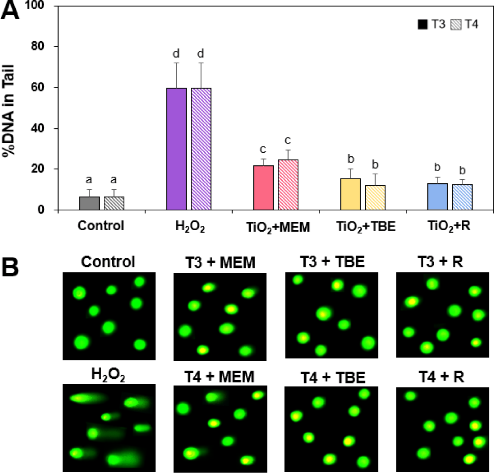
Comet 분석을 이용하여 두 가지 식품첨가물 TiO2 단독처리에 의한 DNA 손상 여부를 평가하였는데, 이는 겔 전기영동 시 손상되어 절단된 DNA 조각들이 핵 외부로 이동하여 혜성 꼬리(comet tail) 모양을 형성하는 원리를 이용하는 기법이다. Comet 분석은 염색체 이상 시험이나 소핵 시험과 달리 DNA 손상을 정량적으로 측정할 수 있어, 초기 유전독성을 평가하는데 유용한 방법이다(Nam et al., 2025). Fig. 5에서 보듯이, 두 가지 식품첨가물 TiO2 단독처리에 의해서 DNA tail이 유의적으로 증가하기는 하나(p<0.05), 양성대조군 H2O2에 의한 DNA tail 생성량보다는 명확히 적은 것으로 나타났다. 선행연구에서 식품용 첨가물 TiO2 (T3 및 T4)가 세포막 손상으로 인한 lactate dehydrogenase 용출이나 세포사멸(apoptosis)을 유발하지 않는 것으로 보고된 바 있으므로(Hwang et al., 2019), 본 연구에서 관찰된 DNA 손상은 apoptosis에 따른 2차적 DNA 단편화가 아닌 TiO2에 의한 직접적인 손상 결과임을 시사한다. TiO2의 광촉매 특성으로 인한 ROS 생성과 이로 인한 세포수준 DNA 손상은 이미 보고된 바 있다(Jeong et al., 2025; Nam et al., 2025). 한편, TBE와 rutin 존재하에 TiO2에 의한 DNA 손상이 유의적으로 감소하였는데, TiO2에 의한 산화스트레스 유발을 고려하면(Fig. 4C, D), TBE와 rutin의 항산화활성에 기인한 것으로 보인다(Fig. 3, Fig. 4E, F). 즉, TiO2에 의한 DNA 손상이 산화스트레스와 연관되어 있음을 입증한다 할 수 있다. 비록 TiO2에 의한 DNA 손상 정도가 TBE 및 rutin과 병용처리 시 대조군 수준으로 감소하지는 않았지만, 이러한 결과는 TBE의 항산화 기능성을 입증하는 것으로, 섭취 수준 및 기간에 따라 유전독성을 억제할 수 있는 잠재력을 보유하고 있음을 시사한다. TBE의 유전독성 완화 효과를 명확히 입증하기 위해, 다양한 세포주로의 확장 연구와 함께 세포신호전달체계를 포함한 세부 메커니즘 규명 및 생체 내(in vivo) 검증이 추가적으로 요구된다.
fep-30-0-260034-g5
Fig. 5. (A) DNA damage (%) and (B) comet assay images of Caco-2 cells following pre-treatment with TBE or rutin and subsequent exposure to TiO2 particles (T3 and T4). All images were magnified at 20×. Different lowercase letters (a~d) above bars indicate significant differences among different samples treated. TBE, Tartary buckwheat extract; R, rutin trihydrate; MEM, minimum essential medium.
Download Original Figure

요 약

본 연구에서는 식품첨가물 착색제로 사용되고 있는 두 가지 다른 입자분포도의 TiO2 유발 산화스트레스와 이로 인한 잠재적 유전독성을 실질적 섭취량을 기반으로 장관세포주에서 평가하였으며, 항산화 기능성 TBE에 의한 유전독성 완화 효과를 연구하고자 하였다. 그 결과, 입자분포도와 상관없이 TiO2는 세포성장을 저해하지는 않으나 ROS를 생성하여 산화스트레스를 유발하였으며, 이는 궁극적으로 DNA 손상을 유도하는 것으로 확인되었다. TiO2에 의해 유발된 산화스트레스 및 DNA 손상은 rutin과 같은 항산화 성분을 보유한 TBE에 의해 완화되는 것으로 나타났다. 본 연구결과는 항산화 기능성 식품 섭취에 의한 TiO2 유발 산화스트레스 및 유전독성 완화 잠재력을 직접적으로 보여주는 결과로, 식품첨가물 착색제 TiO2의 안전성과 TBE의 항산화 기능성에 대한 기초 정보로 활용될 수 있을 것이다.

Conflict of interests

No potential conflict of interest relevant to this article was reported.

Acknowledgements

This research was supported by a National Research Foundation of Korea (NRF) grant funded by the Korean government (MSIT) (RS-2024-00343763), and partly by a research grant from Seoul Women’s University (2026-0225).

Data availability

Upon reasonable request, the datasets of this study can be available from the corresponding author.

Authorship contribution statement

Conceptualization: Choi SJ.

Data curation: Choi SJ.

Formal analysis: Song YS, Choi SJ.

Methodology: Choi SJ.

Software: Song YS.

Validation: Choi SJ.

Investigation: Song YS.

Writing - original draft: Choi SJ.

Writing - review & editing: Song YS, Choi SJ.

Ethics approval

Not applicable.

References

1.

Appel HM, Govenor HL, D’Ascenzo M, Siska E, Schultz JC. 2001. Limitations of folin assays of foliar phenolics in ecological studies. J. Chem. Ecol. 27: 761-778.

2.

Aubert L, Decamps C, Jacquemin G, Quinet M. 2021. Comparison of plant morphology, yield and nutritional quality of Fagopyrum esculentum and Fagopyrum tataricum grown under field conditions in Belgium. Plants. 10: 258.

3.

Dorier M, Béal D, Marie-Desvergne C, Dubosson M, Barreau F, Houdeau E, Herlin-Boime N, Carriere M. 2017. Continuous in vitro exposure of intestinal epithelial cells to E171 food additive causes oxidative stress, inducing oxidation of DNA bases but no endoplasmic reticulum stress. Nanotoxicology. 11: 751-761.

4.

EFSA Panel on Food Additives and Nutrient Sources added to Food (ANS). 2016. Re-evaluation of titanium dioxide (E171) as a food additive. EFSA J. 14: 4545.

5.

Etacheri V, Di Valentin C, Schneider J, Bahnemann D, Pillai SC. 2015. Visible-light activation of TiO2 photocatalysts: advances in theory and experiments. J. Photochem. Photobiol. C: Photochem. Rev. 25: 1-29.

6.

EUR-Lex. 2008. Regulation (EC) No 1333/2008 of the European Parliament and of the Council of 16 December 2008 on food additives (Text with EEA relevance). Available from: https://eur-lex.europa.eu/eli/reg/2008/1333/oj/eng Accessed Jul. 22, 2026.

7.

EUR-Lex. 2022. Commission Regulation (EU) 2022/63 of 14 January 2022 amending annexes II and III to Regulation (EC) No 1333/2008 of the European Parliament and of the Council as regards the food additive titanium dioxide (E 171) (Text with EEA relevance). Available from: https://eur-lex.europa.eu/eli/reg/2022/63/oj/eng Accessed Jul. 22, 2026.

8.

Food and Drug Administration. 2024. Titanium dioxide as a color additive in foods. 21 CFR 73.575. Available from: https://www.fda.gov/industry/color-additives/titanium-dioxide-color-additive-foods?utm Accessed Jul. 22, 2026.

9.

Food Standards Agency. 2024. FSA 22-06-19a-risk analysis process update. Available from: https://webarchive.nationalarchives.gov.uk/ukgwa/20201010091010/http://www.food.gov.uk/print/pdf/node/9616 Accessed Jul. 22, 2026.

10.

Food Standards Australia New Zealand. 2022. Titanium dioxide as a food additive. Available from: https://www.foodstandards.gov.au/sites/default/files/consumer/foodtech/Documents/FSANZ_TiO2_Assessment_report.pdf. Accessed Jul. 22, 2026.

11.

Gräfe C, Weidner A, v.d. Lühe C, Bergemann C, Schacher FH, Clement JH, Dutz S. 2016. Intentional formation of a protein corona on nanoparticles: serum concentration affects protein corona mass, surface charge, and nanoparticle-cell interaction. Int. J. Biochem. Cell Biol. 75: 196-202.

12.

Health Canada. 2002. State of the science of titanium dioxide (TiO2) as a food additive. Available from: https://publications.gc.ca/collections/collection_2022/sc-hc/H164-341-2022-eng.pdf. Accessed Jul. 22, 2026.

13.

Huda MN, Lu S, Jahan T, Ding M, Jha R, Zhang K, Zhang W, Georgiev MI, Park SU, Zhou M. 2021. Treasure from garden: bioactive compounds of buckwheat. Food Chem. 335: 127653.

14.

Hwang JS, Yu J, Kim HM, Oh JM, Choi SJ. 2019. Food additive titanium dioxide and its fate in commercial foods. Nanomaterials. 9: 1175.

15.

Irshad MA, Nawaz R, Rehman MZU, Adrees M, Rizwan M, Ali S, Ahmad S, Tasleem S. 2021. Synthesis, characterization and advanced sustainable applications of titanium dioxide nanoparticles: a review. Ecotoxicol. Environ. Saf. 212: 111978.

16.

Jeong SM, Nam HN, Choi SJ. 2025. Effects of the interactions between food additive titanium dioxide and matrices on genotoxicity. Int. J. Mol. Sci. 26: 617.

17.

Jha R, Zhang K, He Y, Mendler-Drienyovszki N, Magyar-Tábori K, Quinet M, Germ M, Kreft I, Meglič V, Ikeda K, Chapman MA, Janovská D, Podolska G, Woo SH, Bruno S, Georgiev MI, Chrungoo N, Betekhtin A, Zhou M. 2024. Global nutritional challenges and opportunities: buckwheat, a potential bridge between nutrient deficiency and food security. Trends Food Sci. Technol. 145: 104365.

18.

Jin HR, Yu J, Choi SJ. 2020. Hydrothermal treatment enhances antioxidant activity and intestinal absorption of rutin in Tartary buckwheat flour extracts. Foods. 9: 8.

19.

Jo MR, Yu J, Kim HJ, Song JH, Oh JM, Choi SJ. 2016. Titanium dioxide nanoparticle-biomolecule interactions influence oral absorption. Nanomaterials. 6: 225.

20.

Joo YH, Chung NH. 2019. Properties of rutin and its pharmacological effects. Life Sci. Nat. Resour. Res. 27: 47-60.

21.

Kim YH, Youn SM, Yoo NK, Choi SJ. 2021. Cytotoxicity evaluation of frequently used 7 food enzymes in South Korea at their maximum usage levels. Food Eng. Prog. 25: 205-212.

22.

Lee CC, Shen SR, Lai YJ, Wu SC. 2013. Rutin and quercetin, bioactive compounds from tartary buckwheat, prevent liver inflammatory injury. Food Funct. 4: 794-802.

23.

Lee IL, Lee BH, Eom SH, Oh CS, Kang H, Cho YS, Kim DO. 2015. Antioxidant capacity and protective effects on neuronal PC-12 cells of domestic bred kiwifruit. Kor. J. Hort. Sci. Technol. 33: 259-267.

24.

Li X, Zhang Y, Zhao W, Ren T, Wang X, Hu X. 2024. Extracts from tartary buckwheat sprouts restricts oxidative injury induced by hydrogen peroxide in HepG2 by upregulating the redox system. Foods. 13: 3726.

25.

Łabuz P, Gryboś J, Pietrzyk P, Sobańska K, Macyk W, Sojka Z. 2019. Photogeneration of reactive oxygen species over ultrafine TiO2 particles functionalized with rutin-ligand induced sensitization and crystallization effects. Res. Chem. Intermed. 45: 5781-5800.

26.

Mazahir M, Ahmed A, Ahmad A, Ahmad MS, Khan MA, Manzoor MF. 2022. Extraction and determination of bioactive compounds and antioxidant activity of buckwheat seed milling fractions. Food Sci. Technol. 42: e81721.

27.

Ministry of Food and Drug Safety. 2025. Standards and specifications for food additives. MFDS Notification No. 2025-15. Available from: https://www.mfds.go.kr/brd/m_211/view.do?seq=14883 Accessed Jul. 22, 2026.

28.

Mudie DM, Murray K, Hoad CL, Pritchard SE, Garnett MC, Amidon GL, Gowland PA, Spiller RC, Amidon GE, Marciani L. 2014. Quantification of gastrointestinal liquid volumes and distribution following a 240 mL dose of water in the fasted state. Mol. Pharm. 11: 3039-3047.

29.

Nakamura K, Naramoto K, Koyama M. 2013. Blood-pressure-lowering effect of fermented buckwheat sprouts in spontaneously hypertensive rats. J. Funct. Foods 5: 406-415.

30.

Nam HN, Jeong SM, Kim SB, Choi SJ. 2025. Comprehensive evaluation of the genotoxic potential of food additive titanium dioxide in human intestinal cell systems. Int. J. Mol. Sci. 26: 12026.

31.

Peng Q, Wei XQ, Yang Q, Zhang S, Zhang T, Shao XR, Cai XX, Zhang ZR, Lin YF. 2015. Enhanced biostability of nanoparticle based drug delivery systems by albumin corona. Nanomedicine 10: 205-214.

32.

Ranic M, Knez M, Gurinovic M, Vidovic N, Stamataki E, Karapataki K, Zouaghi A, Yercan M, Gunden C, Atakan P, Péter C, Renáta H, Schaer B, Konstadinos M. 2025. Exploring buckwheat's potential—developing innovative dishes, sensory evaluation, consumer acceptance and market integration in Europe: a cross-country analysis. Front. Sustain. Food Syst. 9: 1673883.

33.

Song Y, Cheng Z, Dong Y, Liu D, Bai K, Jarvis D, Feng J, Long C. 2022. Diversity of Tartary buckwheat (Fagopyrum tataricum) landraces from Liangshan, southwest China: evidence from morphology and SSR markers. Agronomy 12: 1022.

34.

Vieira A, Vital N, Rolo D, Roque R, Gonçalves LM, Bettencourtb A, Silva MJ, Louro H. 2022. Investigation of the genotoxicity of digested titanium dioxide nanomaterials in human intestinal cells. Food Chem. Toxicol. 161: 112841.

35.

Worthing M, Mills A, McKenzie-Gopsill A. 2025. Reduced canopy cover and development compromises weed suppression by buckwheat in cover crop mixtures. Can. J. Plant Sci. 105: 1-5.

36.

Yu J, Hwang JS, Oh MS, Lee SY, Choi SJ. 2018. Antioxidant activity of ethanol extracts from common and tartary buckwheat milling fractions. Korean J. Food Sci. Technol. 50: 549-554.

37.

Zhishen J, Mengcheng T, Jianming W. 1999. The determination of flavonoid contents in mulberry and their scavenging effects on superoxide radicals. Food Chem. 64: 555-559.

38.

Zhu F. 2016. Chemical composition and health effects of Tartary buckwheat. Food Chem. 203: 231-245.