
 PDF Download
 PubReader
 Export Citation
 Email the Author
 Share Facebook
 Share Twitter
 Cited by 0 Article
 Metrics (View:619)








Introduction
Seafood is sensitive to heat and temperature. Due to its softer texture compared to meat and high moisture content, microbial and enzymatic activities occur actively, which can easily lead to deterioration and spoilage during distribution (Koo, 1997). Although seafood distribution is primarily conducted in refrigerated or frozen forms, seafood used as home meal replacements (HMR) is mostly distributed in a refrigerated state, suggesting that additional processing may be required for stable distribution. Food poisoning associated with seafood mainly occurs when consuming raw or undercooked fish or shellfish during the summer, and one of the major causative agents is Vibrio species. As the likelihood of Vibrio occurrence increases with rising seawater temperatures in summer, Jung (2024) emphasized that local governments should conduct weekly inspections from May to October to verify compliance with hygienic handling, storage, and distribution standards to prevent this (Um, 2025).
Vibrio is a Gram-negative, comma-shaped bacillus that is motile and classified as a mesophilic aquatic bacterium. Barbieri et al. (1999) reported that Vibrio is found in organic-rich environments such as coastal areas and aquaculture farms worldwide, and is detected in various marine organisms including coral, fish, mollusks, seaweed, and shrimp (Thompson et al., 2004). Among Vibrio species, the major human pathogens include V. parahaemolyticus, V. vulnificus, and V. cholerae.V. parahaemolyticus is a halophilic, Gram-negative bacterium with a single polar flagellum that causes infection through the consumption of raw seafood (Kang et al., 2011). It does not form capsules or spores but possesses virulence factors such as thermostable direct hemolysin (TDH) and TDH-related hemolysin (TRH), which cause gastroenteritis. It is classified as a pathogenic bacterium due to its function of injecting toxins via the Type III Secretion System (TTSS) (Kim et al., 2023). V. parahaemolyticus is widely distributed in coastal waters globally and is primarily detected in seawater with elevated temperatures during the summer. Infection leads to acute gastroenteritis with a latent period of 12–24 hours, followed by symptoms such as fever, vomiting, abdominal pain, and diarrhea, and typically resolves naturally after 2–3 days. The route of infection is oral transmission through water or food contaminated with feces. After a latent period of 1–3 days, severe watery diarrhea and vomiting may occur, and dehydration caused by these symptoms can be fatal (Cha et al., 2012). The cause of these symptoms is the secretion of enterotoxins that adhere to epithelial cells and induce diarrhea, similar to cholera toxin (CT). Vibrio species are sensitive to heat and are easily inactivated in boiling water (100°C); thus, infection can be prevented by sufficiently cooking seafood. However, since most seafood is distributed in a raw form, it cannot be treated with conventional thermal sterilization methods, and the use of chemical additives is difficult due to the characteristics of fresh products. Therefore, a nonthermal process is required to ensure microbial safety without affecting quality, nutrition, or color. Previous studies reported that irradiation of 0–4.0 kGy on seafood such as oysters and shrimp resulted in a reduction of approximately 6 log CFU/g for the nonthermal sterilization of Vibrio (Jakabi et al., 2003; Mahmoud, 2009). A reduction of 3–5 log CFU/g was reported when 3.0 kGy radiation was applied to loach and mackerel (Acharjee et al., 2014). High-pressure processing has been reported to reduce Vibrio in seafood by 2–7.5 log CFU/g at pressures of 250–600 MPa (Kural et al., 2008; Ma & Su, 2011). However, irradiation is difficult to prefer due to negative consumer perception, and high-pressure processing has economic issues requiring expensive equipment and the potential to affect food texture.
Intense Pulsed Light (IPL) sterilization technology inactivates microorganisms on food surfaces by irradiating a broad spectrum of light ranging from ultraviolet (UV) to near-infrared (NIR) for a short duration. This technology is expected to extend shelf life and distribution periods without affecting food quality (Shin et al., 2010; Park & Shin, 2021). The spectrum of IPL is similar to sunlight, but the intensity emitted instantaneously is approximately 20,000 times stronger than sunlight at sea level. Unlike sunlight, the light emitted from IPL has strong UV wavelengths, making it effective for microbial inactivation. UV wavelengths are divided into UV-C (200–280 nm), UV-B (280–315 nm), and UV-A (315–400 nm). Among these, UV-C is absorbed by microbial DNA and exerts the most significant influence through photochemical effects (Cheigh et al., 2012; Mandal et al., 2020). The microbial inactivation mechanism of IPL consists of photochemical, photothermal, and photophysical effects. The photochemical effect occurs when UV-C wavelengths are absorbed by two adjacent thymine bases in microbial DNA, breaking covalent bonds and forming thymine dimers. These dimers convert to pyrimidine dimers, hindering DNA replication and causing mutations, genetic information damage, and replication failure, thereby inactivating the microorganism. The photothermal effect instantaneously transfers heat to microorganisms on the food surface, causing internal heat penetration and local overheating that ruptures cell membranes or inactivates microorganisms due to photothermal stress. However, this is considered a negative factor as it can affect food quality. Regarding photophysical effects, Takeshita et al. (2003) reported that IPL treatment of Saccharomyces cerevisiae resulted in cell shape and membrane deformation, as well as elution of cell contents and proteins. Ramos-Villarroel et al. (2012) observed cytoplasmic damage in Listeria innocua and Escherichia coli upon IPL treatment, and Macias-Rodriguez et al. (2014) reported structural damage, such as central depression in E. coli cells. These studies suggest that IPL contributes to microbial inactivation through photophysical effects as well. Meanwhile, DNA damaged by UV sterilization can be recovered by the cell repair system under certain conditions. However, IPL sterilization inflicts severe damage on DNA, proteins, and macromolecules through stronger energy than continuous UV, further lowering the possibility of recovery (Mandal et al., 2020).
This study analyzed the inactivation effect of V. parahaemolyticus using nonthermal IPL technology and confirmed the reduction rate according to voltage and distance conditions. Additionally, model constants were calculated and equations were derived using the Double-Weibull model to establish IPL treatment conditions for the prevention of food poisoning caused by Vibrio.
Materials and Methods
Vibrio parahaemolyticus (ATCC 17802, KCTC 2729) used in this experiment was obtained from the Korean Collection for Type Cultures (KCTC). A single colony was inoculated into 20% glycerin stock (Daejung Chemicals & Metals Co., Ltd., Siheung, Korea) and stored frozen at –80°C. For the sterilization experiment, the frozen stock was inoculated into 200 mL of NB liquid medium supplemented with 3% NaCl and pre-cultured in a shaking incubator (CSKI-3, Changshin Science Co., Ltd., Seoul, Korea) at 37°C and 180 rpm for 40 h. The cultured broth was then inoculated at 2% into fresh 200 mL NB medium supplemented with 3% NaCl and cultured for 24 h for use in the experiment. The final cell concentration was approximately 8–9 log CFU/mL, and fresh cultures were prepared for each experiment.
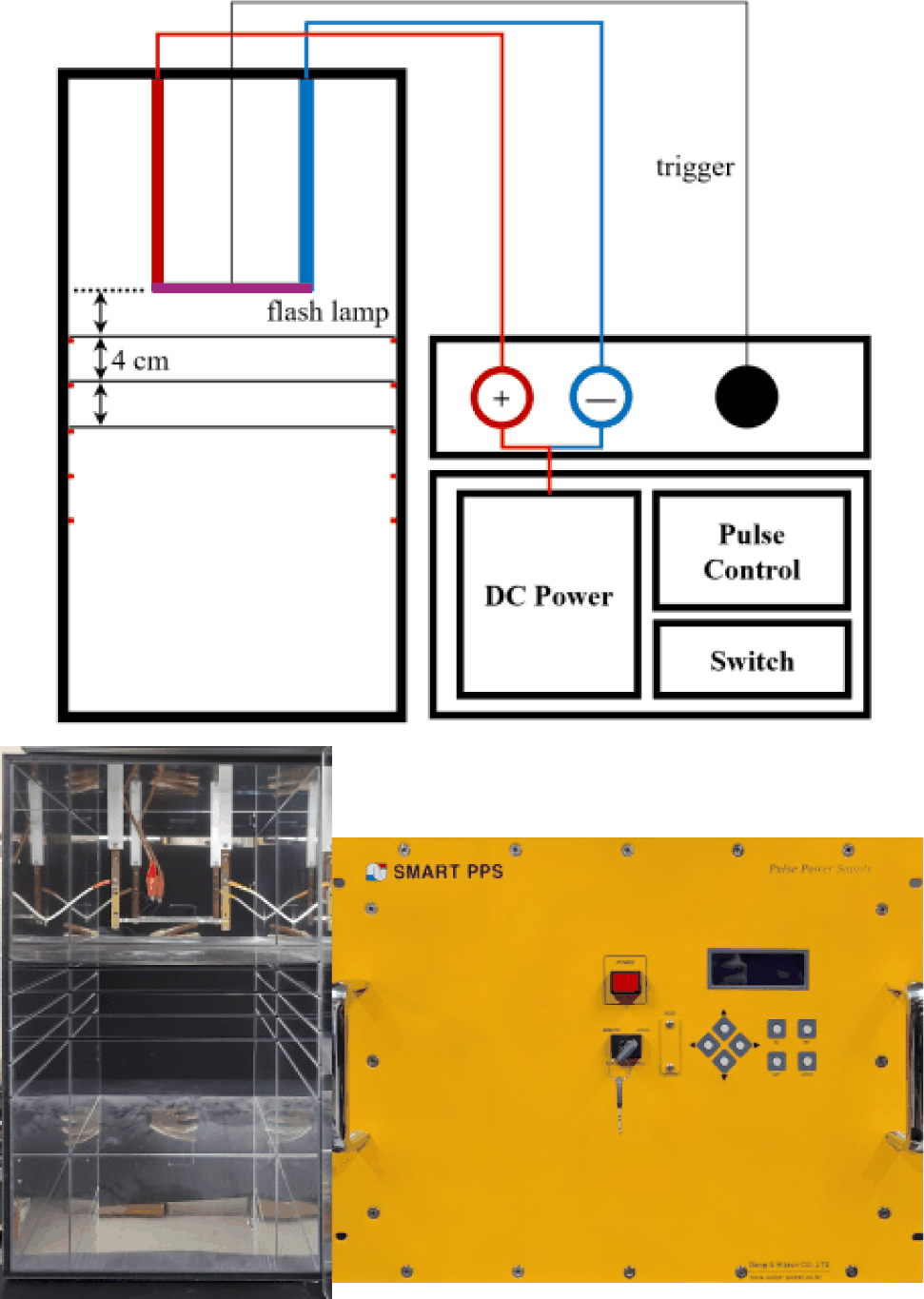
The IPL treatment device used in this experiment consisted of a power supply, pulse generator, light source (lamp), and treatment chamber. The IPL generator (Smart XLPS 2K20Hz, Dong-A Hitech Power Supply, Busan, Korea) used an input power of AC 380 V, 50/60 Hz, and was designed with an average power consumption of 10 kW. The output section was capable of outputting a trigger voltage of 500–2,200 V DC, with a pulse width of 2–500 μs. The current was kept below 1 A for safety. The available frequency was 2–30 Hz. The maximum operation time for a single run was set to 60 min to prevent strain on the device. The treatment chamber was divided to adjust the distance between the light source and the plate, and a reflector was attached inside to minimize light loss. The light source was a xenon XAP series flash lamp (NL 4006, Heraeus Noblelight, Cambridge, UK) filled with mercury-free xenon gas. The overall schematic of the experimental apparatus is shown in Fig. 1. The treatment conditions used in the experiment were voltages of 700–1,000 V, a simmer current of 0.2 A, a pulse width of 20 μs, and distances of 4–12 cm between the light source and the sample. Treatment was performed from 2 to 20 pulses in 2-pulse increments.
To measure the energy density of the light source generated from the xenon lamp during IPL treatment, a radiometer (HD 2102.2, Delta Ohm, Padua, Italy) was used under the same conditions as the experiment. The wavelengths measured by each sensor were divided into UV-C (220–280 nm), UV-B (280–315 nm), UV-A (315–400 nm), and RAD (400–1,050 nm). The energy density for each condition was measured in triplicate, and the average value was calculated.
To check the temperature rise of the plate due to heat generated from the xenon lamp and the influence of the photothermal effect during IPL treatment, the temperature of the plate was measured using a data logger (midi Logger GL200, Graphtec Corp., Yokohama, Japan) under the same conditions used in the experiment. The temperature was measured for 0–4 s at 500 ms intervals, and the minimum value, maximum value, and temperature change were calculated.
The inactivation rate was measured using the spread plate method. The cultured Vibrio broth was centrifuged at 4,000 rpm for 10 min (Gyro 406G, Gyrozen, Daejeon, Korea), washed twice with sterile saline (3% NaCl), and resuspended uniformly. The resuspended culture was serially diluted, and 0.1 mL was spread onto Nutrient Agar (Difco Laboratories) plates supplemented with 3% NaCl. The plates were placed under the xenon lamp of the IPL system and treated with IPL. After treatment, the plates were incubated at 37°C for 24 h, and the number of colonies formed was counted and expressed as CFU/mL. Colony counts between 30 and 300 were used. The inactivation rate of microorganisms (N/N0) was calculated as the ratio of the survival count (N) after treatment to the initial count (N0) and plotted on a semi-log scale. Experiments were repeated 3 times per sample.
To examine the IPL sterilization pattern, the Double-Weibull model proposed by Coroller et al. (2006) was used, and calculations were performed using the GInaFiT tool (version 1.6) developed by Geeraerd et al. (2005). This model is based on the hypothesis that a single population consists of two subpopulations with different resistance to stress, and the inactivation kinetics of the two subpopulations follow a Weibull distribution. The equation of this model is as follows;
Where N0 is the initial cell count, N is the cell count after IPL treatment, and t is the treatment time (μs). α is the difference between the two subpopulations with different inactivation kinetics. δ (δ1, δ2) is the scale parameter, and s is the shape parameter. Here, δ represents the time for the first log reduction, calculated as δ1 and δ2. s indicates the shape of the graph; s>1, s<1, and s=1 represent convex, concave, and linear shapes, respectively.
To test the significance of energy density values and temperature changes according to treatment conditions, Duncan’s multiple range test was used. The significance level for all statistical analyses was p<0.05, and experimental data were measured in triplicate. Statistical analysis was performed using the SPSS Version 28.0 package program (IBM Tech., Chicago, IL, USA).
Results and Discussion
The most critical factor in IPL treatment is the ratio of light wavelengths generated from the xenon lamp, as the broad range of light from ultraviolet to near-infrared affects the sterilization effect. Among the IPL inactivation mechanisms, UV-C in the photochemical effect induces modifications in thymine (T) in the DNA sequence, inhibiting cell replication and proliferation, and its high energy level influences cell inactivation (Alzueta et al., 2019). Additionally, a higher total energy density causes differences in the microbial inactivation rate. The energy density of light reaching the plate during IPL treatment is shown in Table 1. Examining the energy density per pulse according to voltage, the radiation amount for UV-C, UV-B, and UV-A wavelengths tended to increase significantly as the voltage increased. For the energy dose of the UV-C region, it was measured as 3.75 W/m2 at 1,000 V, 3.66 W/m2 at 900 V, 2.87 W/m2 at 800 V, and 2.06 W/m2 at 700 V. On the other hand, the energy dose of the RAD region showed significant differences but no consistent trend, ranging from 13.70 W/m2 at 1,000 V to 13.93 W/m2 at 700 V. Regarding the total radiation amount according to the distance between the lamp and the sample (plate), the radiation amount of all wavelengths tended to decrease as the distance increased. The energy dose of the UV-C region decreased to 3.75 W/m2 at 4 cm, 3.05 W/m2 at 8 cm, and 2.37 W/m2 at 12 cm. The energy dose of the RAD region also decreased from 13.70 W/m2 at 4 cm to 5.50 W/m2 at 12 cm, confirming significant differences according to distance. It is known that the UV-C wavelength range is the most effective for sterilization among various wavelengths in IPL treatment. When the ratio of energy dose of the UV-C region according to voltage is expressed as a percentage, it was 15.69% at 1,000 V, 15.49% at 900 V, 12.74% at 800 V, and 9.98% at 700 V. This predicts that 1,000 V would have high sterilization efficacy due to having the highest energy density as well as the highest UV-C ratio. This difference in ratio due to voltage change can be confirmed, consistent with experiments by Takeshita et al. (2002), Luksiene et al. (2007), and Gómez-López & Bolton (2016). As a result of measuring energy density according to voltage during IPL treatment, sterilization efficacy appeared high in the order of 1,000 V, 900 V, 800 V, and 700 V, which had high total energy density and UV-C wavelength ratios. When the ratio of UV-C wavelength in energy density according to distance was expressed as a percentage (%), it appeared at levels of 15.67% at 4 cm, 18.74% at 8 cm, and 18.73% at 12 cm. The increase in the ratio of UV-C wavelength as the distance increased, despite the same voltage, is thought to be partly influenced by the reflector attached inside the treatment chamber. The sterilization effect according to distance during IPL treatment is predicted to be higher in the order of close distances (4 cm, 8 cm, 12 cm), and similar sterilization effects are expected for similar energy values.
To confirm that IPL operates as a nonthermal processing technology, surface temperature changes were monitored under all experimental conditions (Table 2). The maximum temperature increase (ΔT) observed was 0.85°C (at 1,000 V and 4 cm distance), with most conditions resulting in temperature elevations below 0.50°C. These minimal temperature increases are consistent with the pulsed nature of IPL, where energy is delivered in brief, intermittent bursts rather than continuously (Rowan et al., 1999). The microsecond-duration pulses and low duty cycle (pulse frequency × pulse width = 5 Hz × 20 μs = 0.01%) allow for thermal dissipation between pulses, preventing significant bulk heating (Krishnamurthy et al., 2010). Consequently, heat-sensitive quality attributes of seafood products, including texture, color, flavor, and nutritional components, are expected to be well-preserved during IPL treatment (Ozer & Demirci, 2006). The slightly higher temperature increase observed at 1,000 V and 4 cm distance corresponds to the maximum energy density condition, suggesting a weak correlation between energy input and thermal effects. However, even under these most intensive treatment conditions, the temperature elevation remained well below levels that would induce thermal inactivation of V. parahaemolyticus (typically requiring temperatures exceeding 50°C for significant microbial reduction) (Rippey, 1994).
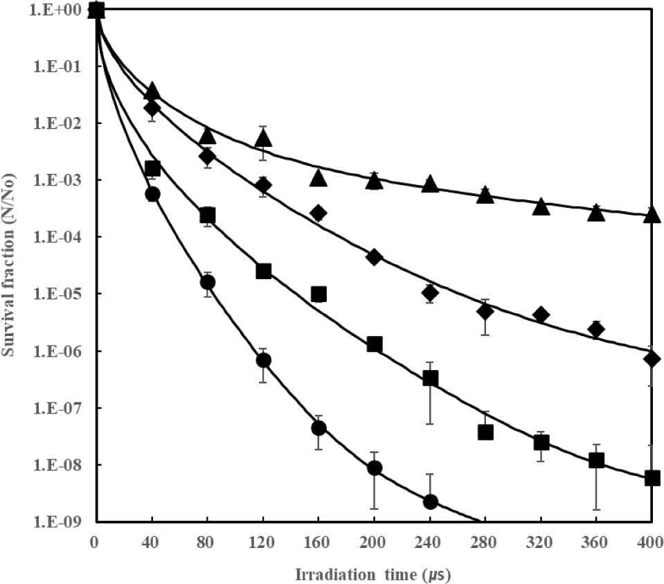
The sterilization effect according to voltage during IPL treatment is shown in Fig. 2. The initial count of V. parahaemolyticus used in the experiment was 9 log CFU/mL. After spreading 0.1 mL on a plate, IPL treatment was performed at a distance of 4 cm from the light source. The treatment time for each voltage was up to the time required for an 8 log reduction or a maximum of 400 μs. Looking at the sterilization effect according to treatment time at 1,000 V, reductions were 3.23 log at 40 μs, 4.79 log at 80 μs, 6.16 log at 120 μs, and 7.34 log at 160 μs. A reduction of more than 8 log was observed when treated for 200 μs or more. At 900 V, a 3.61 log reduction occurred at 80 μs, showing a similar killing effect to 1,000 V at 40 μs, and a 4.59 log reduction at 120 μs was similar to 1,000 V at 80 μs. At 800 V, a 3.09–3.58 log reduction occurred at 120–160 μs, and a 4.35–4.98 log reduction occurred at 200–240 μs. At 400 μs treatment, a 6.14 log reduction was shown. At 700 V, a 3.25 log reduction occurred at 280 μs, showing a similar effect to 1,000 V at 40 μs, and a 3.59 log reduction occurred at 400 μs. The killing effect of V. parahaemolyticus according to voltage showed higher efficacy as the voltage intensity increased, and the effect increased as the treatment time lengthened. The sterilization pattern of V. parahaemolyticus was analyzed using the Double-Weibull model, and the model constants (α, δ1, δ2, s), 4D value (99.99% kill time), and the graph by the model equation are shown in Fig. 2 (R2=0.9836–0.9998) and Table 3. Generally, thermal sterilization shows a linear graph of death rate according to heating time, but nonthermal sterilization methods show various sterilization patterns. In the case of V. parahaemolyticus sterilization by IPL, it showed a curve form indicating resistance after a certain treatment time. The α value by voltage was calculated as 5.20 at 1,000 V, 7.92 at 900 V, 3.60 at 800 V, and 1.84 at 700 V, showing no consistent trend according to voltage. The s value was 0.61 or less at all voltages, appearing as a concave curve graph (s<1). The δ1 value increased as the voltage decreased, being 5.94, 7.20, 17.82, and 19.23 at 1,000 V, 900 V, 800 V, and 700 V, respectively, indicating that the initial sterilization speed was higher as the voltage was higher. However, δ2 did not show a specific trend. Like δ1, the 4D value increased as the voltage decreased, calculated as 58.28 μs at 1,000 V, 94.14 μs at 900 V, 175.15 μs at 800 V, and 556.94 μs at 700 V.
| Voltage (V) | α | s | δ 1 | δ 2 | 4D | R2 |
|---|---|---|---|---|---|---|
| 700 | 1.84 | 0.57 | 19.23 | 144.27 | 556.94 | 0.9836 |
| 800 | 3.60 | 0.61 | 17.82 | 88.21 | 175.15 | 0.9960 |
| 900 | 7.92 | 0.53 | 7.20 | 1,409.10 | 94.14 | 0.9951 |
| 1,000 | 5.20 | 0.61 | 5.94 | 30.46 | 58.28 | 0.9998 |
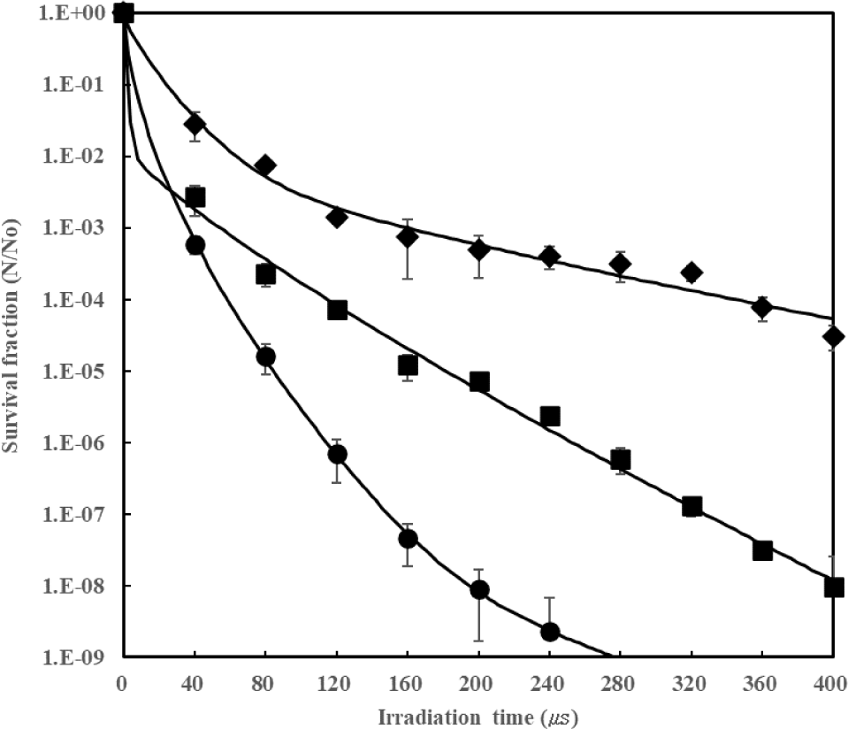
The sterilization effect according to distance during IPL treatment is shown in Fig. 3. The experiment was conducted identically to the voltage effect experiment, treating at distances of 4 cm, 8 cm, and 12 cm at 1,000 V to analyze the sterilization effect over time. The results showed that the closer the distance between the lamp and the plate, the higher the sterilization effect. As the distance increased, the energy density reaching the plate decreased, reducing the sterilization effect. At 4 cm, reductions were 3.23 log at 40 μs, 4.79 log at 80 μs, 6.16 log at 120 μs, 7.34 log at 160 μs, and 8.04 log at 200 μs. At 8 cm, reductions were 3.64 log at 80 μs, 4.92 log at 160 μs, 6.23 log at 280 μs, and 8.02 log at 400 μs, showing a lower sterilization effect than at 4 cm. Meanwhile, at 12 cm, reductions were 3.31 log at 200 μs and 4.52 log at 400 μs, showing significantly lower sterilization effects than at 4 cm and 8 cm. This confirms that the sterilization effect decreases as the distance increases, consistent with the results of Jun et al. (2003), and Sharma & Demirci (2003). The difference in sterilization effect according to the distance between the lamp and the plate becomes clearer when looking at the sterilization effect according to the irradiated energy amount. At 4 cm, a 4.79 log reduction occurred at 95.67 W/m2. At 8 cm, a 4.15 log reduction occurred at 97.36 W/m2. At 12 cm, a 3.12 log reduction occurred at 101.15 W/m2, indicating that more energy was required as the distance increased. Also, looking at the death rate when similar energy amounts were irradiated: at 4 cm, an 8.04 log reduction occurred at 239.18 W/m2; at 8 cm, a 6.23 log reduction at 227.17 W/m2; and at 12 cm, a 4.11 log reduction at 227.58 W/m2, confirming that the death rate decreased significantly as the distance increased. As a result of analyzing the sterilization pattern and model constants according to distance using the Double-Weibull model (Fig. 3 and Table 4), the α value according to treatment time decreased as the distance increased (5.20 at 4 cm, 1.85 at 8 cm, 1.83 at 12 cm). The s value was 0.61 at 4 cm, 0.83 at 8 cm, and 0.80 at 12 cm, appearing as a concave graph (s<1) with no significant trend according to distance. The δ1 value was 5.94 at 4 cm, 2.08 at 8 cm, and 23.95 at 12 cm, showing no trend according to distance. On the other hand, the 4D value was confirmed as 58.28 μs at 4 cm, 114.53 μs at 8 cm, and 343.89 μs at 12 cm. This means that the sterilization effect lowers as the distance increases. The analysis of irradiated energy amount according to distance showed similar results; the 4D energy value was 69.67 W/m2 at 4 cm, 92.46 W/m2 at 8 cm, and 214.29 W/m2 at 12 cm, indicating that the irradiated energy amount must increase as the distance increases to expect a similar sterilization effect. These results indicate that the distance between the lamp and the plate in IPL treatment significantly affects energy density and sterilization efficacy.
| Distance (cm) | α | s | δ 1 | δ 2 | 4D | R2 |
|---|---|---|---|---|---|---|
| 4 | 5.20 | 0.61 | 5.94 | 30.46 | 58.28 | 0.9998 |
| 8 | 1.85 | 0.83 | 2.08 | 45.71 | 114.53 | 0.9918 |
| 12 | 1.83 | 0.80 | 23.95 | 133.43 | 343.89 | 0.9861 |
Conclusion
The application of intense pulsed light (IPL) has been proven as a highly effective nonthermal sterilization strategy for eradicating V. parahaemolyticus in seafood processing. By systematically manipulating operational parameters, this study established that maximizing the voltage to 1,000 V concurrently optimizes the total energy density and elevates the UV-C proportion to 15.69%, while strictly maintaining a surface temperature increase of less than 1°C. This precise balance ensures potent photochemical antimicrobial action without compromising the quality of heat-sensitive seafood. Consequently, a rapid and substantial pathogen reduction exceeding 8 log CFU/mL was accomplished within a mere 200 μs at a close proximity of 4 cm. Furthermore, the successful fitting of the inactivation kinetics to the Double-Weibull model (R2>0.98) demonstrated that the 4D value—the time required for a 99.99% microbial reduction—can be drastically shortened to 58.28 μs under optimal parameters. Ultimately, these robust kinetic analyses and empirical findings provide compelling evidence that IPL technology can be successfully implemented in the seafood industry as a reliable, rapid, and quality-preserving intervention to prevent Vibrio-related foodborne illnesses.


