
Food Engineering Progress
Korean Society for Food Engineering
Reaserch article

생강의 유효성분인 진저론, 6-진저롤 및 6-쇼가올의 발모 촉진 효능 평가

반츠라크치도르젬베1,2,†https://orcid.org/0000-0002-2042-1318, 김진우3,†https://orcid.org/0000-0001-8651-6021, 김진철1,3,*https://orcid.org/0000-0002-2535-2761
Banzragch Dorjsembe1,2,†https://orcid.org/0000-0002-2042-1318, Jin-Woo Kim3,†https://orcid.org/0000-0001-8651-6021, Jin-Chul Kim1,3,*https://orcid.org/0000-0002-2535-2761
1천연물응용과학 과학기술연합대학원대학교
2스마트팜융합연구센터 한국과학기술연구원
3천연물유효성최적화연구센터 한국과학기술연구원
1Natural Product Applied Science, University of Science and Technology (UST), Gangneung 25451, Korea
2Smart Farm Research Center, Korea Institute of Science and Technology (KIST), Gangneung 25451, Korea
3Center for Natural Product Efficacy Optimization, Korea Institute of Science and Technology (KIST), Gangneung 25451, Korea
*Corresponding author: Jin-Chul Kim, Natural Product Applied Science, University of Science and Technology (UST), Gangneung 25451, Korea, E-mail: jckim@kist.re.kr

These authors contributed equally to this work.

ⓒ Copyright 2026 Korean Society for Food Engineering. This is an Open-Access article distributed under the terms of the Creative Commons Attribution Non-Commercial License (http://creativecommons.org/licenses/by-nc/4.0/) which permits unrestricted non-commercial use, distribution, and reproduction in any medium, provided the original work is properly cited.

Received: Aug 18, 2026; Revised: Sep 15, 2026; Accepted: Oct 02, 2026

Published Online: Oct 27, 2026

Abatract

Natural therapeutic agents have attracted considerable attention as potential treatment options for alopecia. This study compared the hair growth-promoting effects of Zingiber officinale compounds (zingerone, 6-gingerol, and 6-shogaol) on human dermal papilla cells (HDP cells) and C57BL/6 mice. In vitro, all three compounds increased the expression of phosphorylated Akt (p-Akt), phosphorylated extracellular signal-regulated kinase (p-ERK), Sonic Hedgehog (SHH), suppressor of Fused (SUFU), and Cyclin D3 in HDP. However, in vivo, only 6-gingerol significantly promoted hair regeneration and induced skin darkening. Histological analysis confirmed that 6-gingerol facilitated telogen-to-anagen transition, extending the hair bulbs into the deep subcutis. qRT-PCR showed that 6-gingerol markedly upregulated key Wnt/β-catenin components (β-catenin, LEF1, Wnt5a) and VEGF in skin tissues, whereas zingerone failed to activate β-catenin/LEF1 and 6-shogaol downregulated most hair growth genes. In conclusion, 6-gingerol is the primary active component of Z. officinale that induces hair growth by activating the Wnt/β-catenin and VEGF pathways.

Keywords: Zingiber officinale; Hair growth; Dermal papilla cells; Wnt/β-catenin pathway

서 론

모발은 체온 조절, 두피 보호 및 유해 물질 배출과 같은 생체 방어 기능뿐만 아니라(Kinde et al., 2024), 개인이 타인에게 보이는 외형적 이미지를 형성하여 자아존중감과 사회적 관계 형성에 큰 영향을 미친다(Dhami, 2021). 모발은 성장이 일어나는 생장기(anagen), 모낭이 퇴화하는 퇴행기(catagen) 및 모발이 탈락하는 휴지기(telogen)의 규칙적인 순환 과정을 거치며, 지속적으로 재생과 탈락을 반복하는 모발 주기를 가진다(Lin et al., 2022). 특히 휴지기에서 생장기로의 전환을 비롯한 모발 주기 관리는 모낭 기저부의 모유두세포가 담당한다(Wu et al., 2021). 모유두세포는 모낭의 초기 형성, 크기 결정 및 생장기 유지를 총괄하는 핵심 세포군으로(Morgan, 2014), 모유두세포의 증식은 대표적인 세포사멸 억제 및 생존 신호전달 경로인 Akt와 ERK의 활성화(Han et al., 2004), 세포주기 진행 단백질인 Cyclin D의 발현 촉진(Rastegar et al., 2015), 그리고 모낭의 재생 및 생장기 전환을 결정지어 주는 Wnt/β-catenin과 Sonic Hedgehog (SHH) 신호전달 경로의 작동에 의해 종합적으로 유도된다(Choi, 2020). 또한, 모유두세포에서 분비되는 VEGF, HGF, IGF 등의 성장인자는 모낭 주변의 미세혈관 형성 및 영양 공급을 촉진함으로써 모발의 성장을 지속적으로 유지시킨다(Qi et al., 2016; Choi, 2020).

탈모(alopecia)는 이러한 모발 주기에 이상이 생겨 발생하는 질환으로, 모발이 존재해야 할 부위에 정상적으로 존재하지 않거나 과도한 탈락이 일어나는 상태를 의미한다. 탈모는 과거 주로 유전적 요인이나 연령 증가에 의해 발생하는 것으로 알려졌으나(Paus & Cotsarelis, 1999), 최근 스트레스, 환경오염, 식습관 변화 및 호르몬 불균형 등 다양한 원인으로 인해 연령과 성별에 관계없이 발병 빈도가 증가하는 추세이다(Sankhwar & Khan, 2025). 탈모는 치명적인 신체적 장애를 일으키지는 않으나, 환자의 외형적 변화에 따른 심리적 불안감과 사회적 위축을 초래하여 정신 건강과 삶의 질을 크게 저하시킨다(Cash, 1992).

현재 탈모의 치료 및 완화를 위해 사용되는 대표적인 약품으로는 FDA 승인 의약품인 미녹시딜이 있다(Gupta et al., 2022). 미녹시딜은 두피 미세혈관을 확장함과 동시에, 모유두세포 내 Akt 및 ERK 신호전달 경로를 활성화하여 세포 증식을 유도하고 VEGF 등 성장인자의 분비를 촉진하는 것으로 알려져 있다(Han et al., 2004). 하지만, 미녹시딜은 두피 자극, 가려움증, 홍반 및 전신 혈압 저하 등의 부작용이 동반될 수 있어(Suchonwanit et al., 2019), 부작용이 적고 장기적으로 적용할 수 있는 천연물 기반 발모 소재가 각광받고 있는 추세이다(Natarelli et al., 2023; Paik et al., 2025).

생강(Zingiber officinale Rosc.)은 전통적으로 식용 및 약용으로 널리 사용되어 온 생강과 식물로, 항산화, 항염증 및 혈액순환 개선 등 다양한 생리활성을 갖는 것으로 알려져 있다(Mao et al., 2019). 최근 생강 추출물 및 유효성분의 발모 촉진 가능성이 보고되고 있으나(Abbas, 2020; Putra et al., 2020; Li et al., 2024), 상반되는 결과를 가진 연구도 존재하여(Miao et al., 2013; Lie et al., 2024) 생강 및 유효성분의 발모 효능이 명확히 규정되지 않은 실정이다. 특히, 생강의 대표 지표 성분인 6-gingerol은 열처리 및 건조 가공 시 탈수 반응에 의해 6-shogaol로 전환되며, 고온 조건의 열분해 과정을 거쳐 zingerone으로 분해되는 특성을 지닌다(Bhattarai et al., 2001). 따라서 생강의 가공 조건에 따라 이들 성분의 조성 비율이 크게 달라질 수 있어 각 성분의 명확한 효능 평가가 필요함에도 불구하고, 지금까지 이들 단일 성분 간의 효능을 직접 비교한 연구는 미흡한 실정이다. 따라서 본 연구에서는 생강의 주요 유효 성분(6-gingerol, 6-shogaol, zingerone)의 발모 조절 효능을 in vitro 및 in vivo 모델에서 평가함으로써, 각 성분별 작용 특성과 관련 기전을 규명하고 식품 유래 발모 촉진 소재로서의 활용 가능성을 검증하고자 하였다.

재료 및 방법

윤리 승인

동물실험은 한국과학기술연구원 동물실험윤리위원회(Institutional Animal Care and Use Committee, IACUC)의 승인을 받았으며(승인번호: No. 2018-093), 미국 국립보건원(NIH)에서 발행한 실험동물 관리 및 사용 지침(NIH 간행물 No. 85-23, 1996년 개정)에 따라 수행되었다.

실험 재료 및 시약

본 연구에 사용된 zingerone, 6-gingerol, 6-shogaol 및 미녹시딜(minoxidil, MXD) Sigma-Aldrich사(St. Louis, MO, USA)로부터 구입하여 사용하였다. 또한, 조직학적 분석을 위한 헤마톡실린(hematoxylin), 에오신(eosin), 자일렌(xylene), 10% 중성 완충 포르말린(10% neutral buffered formalin) 및 에탄올(ethanol, EtOH) 역시 Sigma-Aldrich사에서 구입하여 분석에 사용하였다.

인간모유두세포 배양

인간모유두세포(Human Dermal Papilla Cells, HDP 세포)는 바이오드림(BioDream, Chuncheon, Korea)에서 분양받았으며, 배지는 제조사에서 제공하는 CEFOgro™ Human Dermal Papilla Growth Medium 500 mL에 동봉된 첨가제(supplements) 20 mL 및 페니실린-스트렙토마이신(penicillin-streptomycin) 2.5 mL을 첨가하여 사용하였다. 세포는 100 mm cell culture dish에 seeding하고 37℃, 5% CO2 incubator에서 배양하였다. 배지는 2일에 한 번씩 교환하였으며, cell confluence가 80~90% 정도가 되면 0.05% Trypsin-EDTA를 이용하여 계대배양을 수행하였다.

세포 생존율 측정

Zingerone, 6-gingerol 및 6-shogaol이 HDP 세포의 생존율에 미치는 영향을 평가하기 위해, MTT assay를 수행하였다. 96-well 플레이트에 2×10⁴ cells/well 농도로 HDP 세포를 분주한 뒤, 37℃, 5% CO2 조건 하에서 24시간 동안 배양하였다. 이후 각 well에서 배지를 제거하고, 각 시험 물질을 농도별(0, 5, 10, 20 및 40 µM)로 희석한 배지 100 µL를 각 well에 첨가하여 추가로 37℃, 5% CO2 조건 하에서 반응시켰다. 노출이 끝난 후 배지를 버리고, 0.5 mg/mL 농도의 MTT 시약이 첨가된 배지 100 µL를 분주하여 1시간 동안 반응을 유도하였다. 생성된 formazan 침전물을 용해하기 위해 MTT 반응액을 제거한 뒤 100 µL의 DMSO를 가하였다. 최종적으로 마이크로플레이트 분광광도계(Infinite M1000, TECAN Ltd., Männedorf, Switzerland)를 사용하여 560 nm 파장에서 흡광도를 측정하였다.

Western blot

60 mm cell culture dish에 1×10⁵ cells/dish 농도로 HDP 세포를 분주한 후, 37℃, 5% CO2 조건 하에서 24시간 동안 배양하였다. 배양 종료 후 dish의 배지를 제거하고 supplement가 포함되지 않은 배지로 교체하여 24시간 동안 starvation을 수행하였다. 이후 배지를 다시 제거하고, supplement가 없는 배지에 각 sample (10 µM)을 용해하여 처리한 뒤 24시간 동안 배양하였다. 배양이 끝난 뒤, dish의 배지를 제거한 뒤, DPBS를 이용하여 3회 세척하였다. 이후, RIPA buffer (ThermoFisher Scientific, IL, USA)를 이용하여 세포를 용해한 뒤, 13,000 rpm, 4℃ 조건으로 15분간 원심분리하였다. 이어서, 상층액을 취한 뒤, BCA 단백질 정량 키트(BCA protein assay kit; Thermo Scientific, Cleveland, OH, USA)를 사용하여 단백질 농도를 측정하였다. 각 시료당 10 µg의 단백질을 SDS-PAGE (sodium dodecyl sulfate-polyacrylamide gel electrophoresis)로 분리한 뒤 PVDF (polyvinylidene difluoride) membrane (Millipore, Billerica, MA, USA)으로 전이(transfer)시켰다. 단백질이 전이된 membrane은 5% BSA (bovine serum albumin)가 포함된 PBS 용액을 이용하여 25℃에서 1시간 동안 blocking하였다. 이후 membrane은 5% BSA에 1:1,000으로 희석한 1차 항체를 첨가하여 4℃에서 15시간 동안 반응시켰다. 1차 항체로는 p-Akt, Akt, p-ERK, ERK, SHH, SUFU, cyclin D3을 사용하였으며, loading control로 GAPDH를 사용하였다. 이후 membrane을 5% BSA에 1:10,000 비율로 희석한 2차 항체와 25℃에서 1시간 동안 반응시켰다. 이어서 SuperSignal™ West Femto Maximum Sensitivity Substrate (Thermo Fisher Scientific, Waltham, MA, USA)를 처리하여 신호를 증폭시킨 후, iBright CL1000 system (Thermo Fisher Scientific)을 통해 결과를 분석했다. 본 연구에 사용된 모든 항체에 대한 세부내용은 Table 1에 기입하였다.

Table 1. Antibodies for western blot analysis
Target Host species / type Dilution rate Brand Cat. no.
p-Akt Rabbit IgG / primary 1:1,000 CST 4060
Akt Rabbit IgG / primary 1:1,000 CST 4685
p-ERK Rabbit IgG / primary 1:1,000 CST 4370
ERK Rabbit IgG / primary 1:1,000 CST 4695
SHH Rabbit IgG / primary 1:1,000 CST 2207
SUFU Rabbit IgG / primary 1:1,000 CST 2520
Cyclin D3 Rabbit IgG / primary 1:1,000 CST 2936
GAPDH Rabbit IgG / primary 1:10,000 CST 2118
Rabbit IgG Mouse IgG-HRP /secondary 1:10,000 SCBT SC-2357

p-Akt, phosphorylated protein kinase B; p-ERK, phosphorylated extracellular signal-regulated kinase; SHH, sonic hedgehog; SUFU, suppressor of fused homolog; GAPDH, glyceraldehyde 3-phosphate dehydrogenase; IgG, immunoglobulin G; HRP, horseradish peroxidase. CST, Cell Signaling Technology (Danvers, MA, USA); SCBT, Santa Cruz Biotechnology (Dallas, TX, USA).

Download Excel Table
실험동물군 설정 및 실험물질 도포

8주령 수컷 C57BL/6 마우스는 OrientBio (Seongnam, Korea)에서 구입하여, 무병원성 동물 시설에서 6일간 순화하였다. 시설은 22±2℃의 온도와 50±10%의 습도를 유지하고, 12시간 명암 주기로 관리되었으며, 마우스는 실험 기간 동안 표준 사료와 물을 자유롭게 섭취하였다. 순화 후 마우스의 등은 제모기를 사용하여 1차로 털을 제거하였으며, 제모제를 사용하여 잔여 털을 제거하였다. 이후, 하루간 추가 순치 과정을 거쳤다. 이후 마우스는 무작위로 5개의 군(n=5)으로 나누었다: 대조군(control, CON), 75% 에탄올 처리; MXD 처리군(양성 대조군), 5% 미녹시딜 처리; zingerone 처리군, 75% 에탄올에 녹인 zingerone (1 mg/mL) 처리; 6-gingerol 처리군, 75% 에탄올에 녹인 6-gingerol (1 mg/mL) 처리; 6-shogaol 처리군, 75% 에탄올에 녹인 6-shogaol (1 mg/mL) 처리. 이때 시험 물질의 도포 농도(1 mg/mL)는 마우스 등 피부 국소 도포 시 피부 자극이나 이상 징후를 유발하지 않고 피부 생리활성 효능이 보고된 생강 활성 성분의 선행 연구(Wu et al., 2010; Meena et al., 2025)를 참고하여 설정하였으며, 세 성분 간의 효능을 동일 조건에서 비교하기 위해 동일 농도로 적용하였다. 시료는 55일간 매일 동일한 시간에 100 µL씩 마우스의 오른쪽 등에 도포하였다. 실험 기간 동안 주기적으로 마우스 등 부위를 사진 촬영하였으며, 55일차에 마우스를 희생시켜 등 피부 조직을 채취하였다. 마우스 등 발모 정도는 독립된 평가자 2인이 무작위 맹검(double-blind) 방식으로 평가하였다. 발모 점수(hair growth score)는 등 피부의 색조 변화 및 털의 생장 상태에 따라 0점(분홍색 피부, 발모 징후 없음), 1점(회색 피부, 초기 발모 징후), 2점(짙은 회색 피부, 발모 진행), 3점(검은색 피부, 모발의 완전한 성장)으로 차등 부여하였으며(Table 2), 두 평가자의 점수를 평균하여 최종 분석에 활용하였다.

Table 2. Scoring of hair growth and criteria for grading
Hair growth score Observation
0 Pink skin (no hair growth)
1 Gray skin (initial hair growth)
2 Darker gray (indicative of more advanced hair growth)
3 Black (fully grown hair)
Download Excel Table
헤마톡실린-에오신(H&E) 염색

적출한 마우스의 등 피부 조직은 10% 중성 완충 포르말린 용액에 24시간 동안 고정한 후, 파라핀에 포매하였다. 포매된 블록은 박절기(microtome)를 이용하여 5 µm 두께로 조직 절편을 제작하였다. 이후 자일렌에 10분씩 3회 침지하여 탈파라핀화를 진행하였으며, 에탄올(100%, 95%, 70%, 50%) 수용액을 각각 3분간 순차적으로 침지시켜 조직을 수화시켰다. 피부의 병리학적·형태학적 변화를 관찰하기 위해 H&E 염색을 실시하였다. 염색이 완료된 조직 절편은 광학현미경(Olympus CX43, Tokyo, Japan) 및 Cellsens Standard 소프트웨어를 통해 관찰하였으며, 현미경용 카메라 시스템(TE-2000U, Nikon Instruments Inc., Melville, USA)을 활용하여 조직 이미지를 획득하였다.

유전자 발현 비교 분석

마우스 등 조직을 Biomasher (Nippi, Tokyo, Japan)로 균질화한 후, RNeasy® Mini Kit (Qiagen, Hilden, Germany)으로 총 RNA를 추출하였다. 추출된 RNA는 RevertAid First Strand cDNA Synthesis Kit (Thermo Scientific, MA, USA)로 cDNA로 역전사하였다. qRT-PCR은 Power SYBR Green Master Mix (ThermoFisher Scientific)를 사용하여 AP7500 qRT-PCR System (ThermoFisher Scientific)에서 수행하였다. PCR 조건은 초기 단계(50℃ 2분, 95℃ 20초) 후, 40 사이클(95℃ 20초, 57℃ 1분)로 진행하였다. Gapdh를 housekeeping gene으로 사용하여 표적 유전자 발현을 정규화하였다. 상대 발현은 2-ΔΔCT 방법으로 계산하였으며(Livak & Schmittgen, 2001), 사용된 프라이머(primer) 정보는 Table 3에 기재하였다.

Table 3. Primer sequences for qRT-PCR
Target (genetic symbol) Sequences (5'-3')
VEGF (Vegfa) Forward CACGACAGAAGGAGAGCAGA
Reverse CAGGGCTTCATCGTTACAGC
WNT5A (Wnt5a) Forward CTGCTTTGGCAGGGTGATG
Reverse TAGTCGATGTTGTCTCCGCA
β-Catenin (Ctnnb1) Forward ACCCTGGTGCTGACTATCCA
Reverse AGTATCAAACCAGGCCAGCT
LEF1 (Lef1) Forward GGCATCCCTCATCCAGCTAT
Reverse ACTCTCCTTTAGCGTGCACT
HGF (Hgf) Forward TCAAGGCCAAGGAGAAGGT
Reverse ACTCGGATGTTTGGGTCAGT
IGF (Igf1) Forward AGTTTTGTGTTCACCTCGGC
Reverse TGAAGTAAAAGCCCCTCGGT
GAPDH (Gapdh) Forward CATGGCCTTCCGTGTTCCTA
Reverse ACTTGGCAGGTTTCTCCAGG
Download Excel Table
통계 처리

모든 실험 데이터는 IBM SPSS Statistics 21 (IBM Corp., Chicago, IL, USA)을 이용하여 통계 분석을 수행하였으며, 결과는 평균±표준편차(mean±SD)로 표기하였다. 실험 군 간의 유의성 검정은 일원배치 분산분석(one-way ANOVA)을 실시한 후 Dunnett's test를 통해 사후검정을 수행하였다. 통계적 유의성은 p<0.05, p<0.01 및 p<0.001 수준에서 판정하였다.

결과 및 고찰

생강 유효 성분의 모유두세포 내 발모 관련 신호 전달 경로 및 세포 주기 조절 단백질 발현 영향

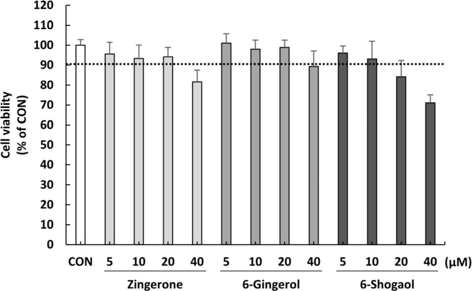
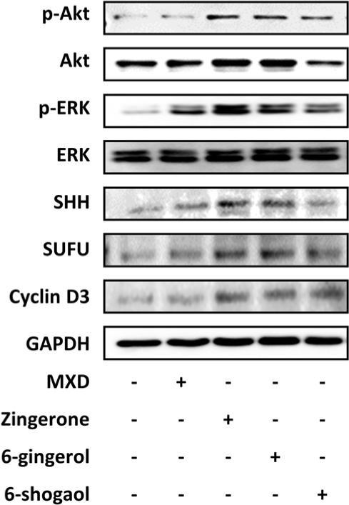
모유두세포는 모낭의 형성과 성장 주기를 조절하는 핵심 세포로, 이들의 증식은 모발 성장의 필수 전제 조건이다(Houschyar et al., 2020). Akt 및 ERK 기전의 활성화는 모유두세포의 세포사멸을 억제하고 증식을 유도하는 대표적인 신호전달계이며(Han et al., 2004), SHH 경로는 모낭의 상피-간엽 상호작용(Epithelial-mesenchymal interaction)을 자극하여 모낭 형성을 유도한다(Choi, 2020). 또한 Cyclin D의 발현 증가가 세포주기 G1/S 진입을 촉진하여 세포의 증식을 유도한다(Rastegar et al., 2015). 생강의 주요 유효 성분인 zingerone, 6-gingerol 및 6-shogaol이 HDP 세포의 발모 관련 신호 전달 경로 및 세포 주기 조절 단백질 발현에 미치는 영향을 확인하고자 Western blot 분석을 수행하였다. 이때 처리 농도는 MTT 평가에서 세 물질 모두 세포 생존율을 90% 이상 안정적으로 유지하여 유의한 독성을 나타내지 않은 10 µM (Fig. 1)로 일괄 결정하여 수행하였다. 실험 결과, zingerone, 6-gingerol 및 6-shogaol 처리군 모두에서 세포 생존 및 증식에 관여하는 p-Akt 및 p-ERK의 발현 수준이 control 대비 현저히 증가하였으며, 양성 대조군인 MXD와 비등하거나 이를 상회하는 높은 활성화를 보였다(Fig. 2). SHH 경로를 분석한 결과, SHH 및 SUFU 단백질의 발현이 zingerone 및 6-gingerol 처리군에서 뚜렷하게 증가함을 확인하였다. 또한, 생강 유효 성분 3종 처리군 모두에서 control 대비 Cyclin D3의 발현이 유의하게 증가하였다(Fig. 2). 따라서, zingerone, 6-gingerol 및 6-shogaol은 in vitro 수준에서 Akt, ERK 및 SHH 신호 전달 경로의 활성화와 Cyclin D3의 발현을 증가시켜 모유두세포의 증식을 촉진하고 발모를 유도하는 것으로 판단된다.
fep-30-0-260038-g1
Fig. 1. Effects of zingerone, 6-gingerol, and 6-shogaol on the cell viability of HDP cells. Data are presented as mean±standard deviation (SD).
Download Original Figure
fep-30-0-260038-g2
Fig. 2. Effects of zingerone, 6-gingerol, and 6-shogaol on hair growth-related protein expressions in HDP cells. Represented image of western blot data. Protein expression levels of p-Akt, Akt, p-ERK, ERK, SHH, SUFU, and Cyclin D3 were evaluated by Western blot after treatment with MXD, Zingerone, 6-gingerol, and 6-shogaol in HDP cells.
Download Original Figure
생강 유효 성분의 등 피부 국소 도포에 따른 마우스 발모 촉진 및 재생 효능 평가

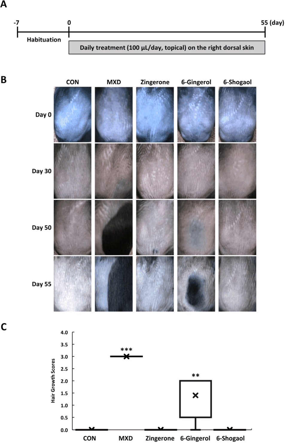
In vivo 모델에서 zingerone, 6-gingerol 및 6-shogaol의 실질적인 발모 촉진 효능을 평가하고자 하였다. 이때, 자연적인 발모주기 변수를 통제하고 모낭의 휴지기 동기화를 유도하기 위해 8주령 수컷 C57BL/6 마우스를 사용하였다(Müller-Röver et al., 2001). 제모된 마우스의 등 부위에 생강 유효 성분을 매일 국소 도포하여 경과에 따른 발모 촉진 효과를 육안 관찰 및 발모 점수 분석을 통해 평가하였다(Fig. 3A). 휴지기 상태의 C57BL/6 마우스 피부는 핑크색을 나타내지만, 모낭이 생장기로 진입함에 따라 모구(hair bulb) 부위의 멜라닌 합성으로 피부색이 짙은 검은색으로 전환된다. 따라서 육안으로 털이 관찰되기 전 피부가 흑색으로 변하는 것은 모낭이 생장기 단계로 진입했음을 보여주는 주요 지표로 활용된다(Slominski & Paus, 1993; Hwang et al., 2022). 실험 결과, control, zingerone 및 6-shogaol 처리군은 관찰 기간 동안 등 피부의 색조 변화 및 모발의 재생이 전혀 관찰되지 않았다. 반면, 양성 대조군인 MXD 처리군에서는 30일차에 피부색이 짙은 검은색으로 변화한 후, 모발이 성장함을 확인하였다. 생강 유효 성분 중 6-gingerol 처리군은 50일차에 도포 부위를 중심으로 피부 착색 및 뚜렷한 모발 재생 영역이 유의미하게 증가하며, 55일차에는 털이 자라나는 것을 확인하였다(Fig. 3B). 이를 정량적으로 평가하기 위해, Hair growth score를 활용하여 발모 촉진정도를 확인하였다. 55일차에, control, zingerone 및 6-shogaol 처리군은 score가 0으로 발모가 전혀 진행되지 않았다. 반면, MXD 처리군은 3.0을 나타내어 발모가 충분히 진행되었으며, 6-gingerol 처리군은 score가 1.4를 나타내어 6-gingerol이 모발 성장 유도 효능이 있음이 입증되었다(Fig. 3C).
fep-30-0-260038-g3
Fig. 3. Effects of zingerone, 6-gingerol, and 6-shogaol on hair growth in C57BL/6 mice. (A) Schematic diagram illustrating the experimental design and treatment schedule. (B) Representative photographs of the dorsal skin from each group at days 0, 30, 50, and 55. (C) Quantitative evaluation of hair growth score on day 55. Groups: CON (75% EtOH), MXD (5% minoxidil), zingerone, 6-gingerol, and 6-shogaol (1 mg/mL in 75% EtOH). Data are presented as a box-and-whisker plot, where 'x' indicates the mean value. Statistical significance was determined using one-way ANOVA followed by Dunnett's post-hoc test (**p<0.01 and ***p<0.001 vs. CON).
Download Original Figure
생강 유효 성분의 마우스 피부 조직 내 모낭 발달 및 조직학적 변화 평가

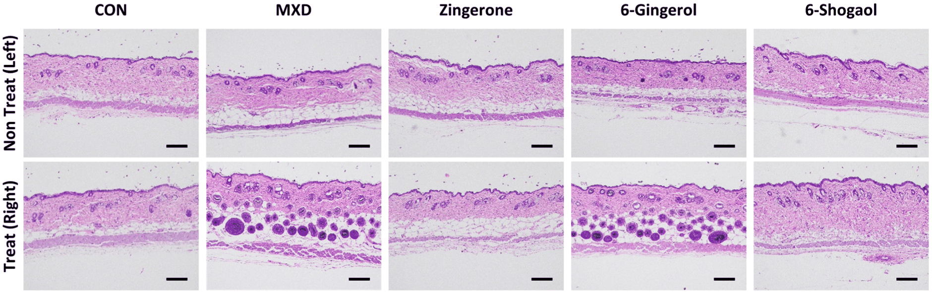
모발 성장 주기 변화는 피부 두께와 모낭의 공간적 위치 변화를 동반한다(Paus & Cotsarelis, 1999). 발모가 촉진되면 피부 두께는 가장 두꺼워지며 모구가 진피 하부 및 피하 지방층 깊숙이 확장된 생장기 모낭 구조로 전환된다. 반면, 휴지기에는 피부가 가장 얇아지며 모낭의 위치가 진피층으로 올라오게 된다(Müller-Röver et al., 2001). 생강 유효 성분인 zingerone, 6-gingerol 및 6-shogaol의 도포가 마우스 등 피부 조직의 모낭 구조 및 형태학적 변화에 미치는 영향을 확인하기 위해 피부 조직 절편에 대한 H&E 염색 후, 분석을 수행하였다. 조직 염색 결과, 물질을 처리하지 않은 무처리 부위는 control과 마찬가지로 조직학적 변화가 관찰되지 않았다(Fig. 4). Control을 포함하여 zingerone 및 6-shogaol 처리군 역시 모낭의 크기가 작고 주로 진피 상부에 머물러 있었다. 반면, MXD 및 6-gingerol 처리군에서는 모낭의 수와 크기가 확연히 증가하였을 뿐만 아니라, 모구가 진피 하부 및 피하 지방층 깊숙이 확장된 것을 확인하였다(Fig. 4).
fep-30-0-260038-g4
Fig. 4. Effects of zingerone, 6-gingerol, and 6-shogaol on hair follicle development in mouse skin tissues. Hematoxylin and eosin (H&E) stained dorsal tissue sections from mice in each group. The top panel shows the normal skin area without any treatment, and the bottom panel shows the skin area topically treated with each compound (scale bar: 100 µm).
Download Original Figure
생강 유효 성분의 모발 성장 관련 유전자발현에 미치는 영향

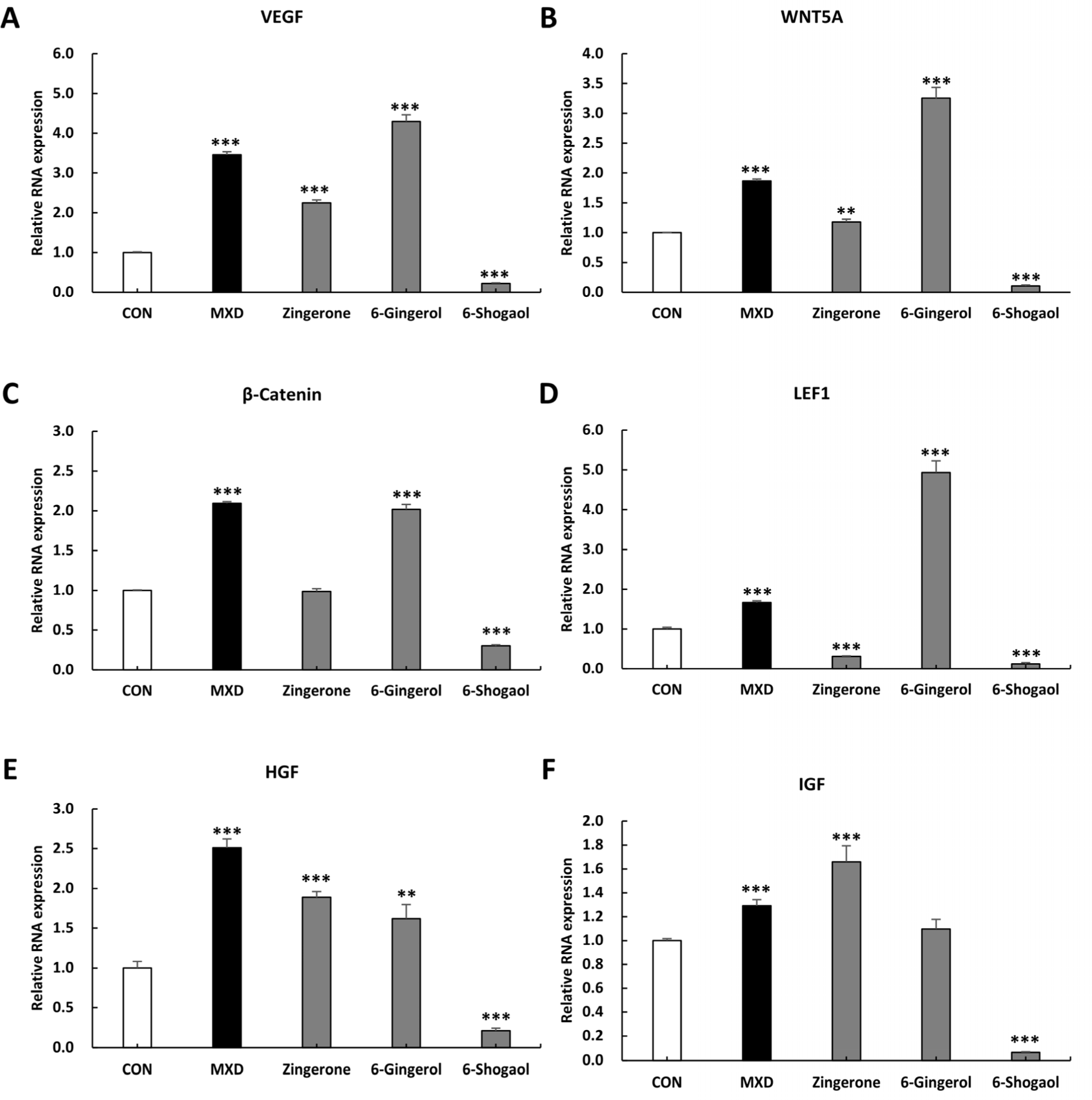
피부 조직 내 모발 성장 과정에서 β-catenin 및 LEF1은 모낭 줄기세포의 활성화와 휴지기 모낭의 생장기 진입을 유도하는 Wnt 신호전달계의 필수 전사 인자이며(Choi, 2020), Wnt5a는 모낭의 구조적 형태 형성 및 세포 분화를 조절한다(Reddy et al., 2001). 또한 VEGF는 모낭 주변 미세혈관 형성을 통한 영양 공급을 촉진하고(Yano et al., 2001), HGF와 IGF는 모유두세포의 증식을 자극하고 모발 생장기를 유지시키는 주요 성장인자이다(Zhang et al., 2024; Kwack et al., 2026). 피부 조직 내 모발 성장 관련 유전자 발현을 qRT-PCR로 분석한 결과, VEGF 및 Wnt5a의 발현은 control 대비 MXD, zingerone 및 6-gingerol 처리군에서 유의하게 증가하였으나, 6-shogaol 처리군에서는 오히려 감소하였다(Fig. 5A, B). 모낭 생장기 전환의 핵심 전사인자인 β-catenin의 발현은 control 대비 MXD 및 6-gingerol 처리군에서만 유의하게 증가하였으며, zingerone 처리군은 유의적 변화가 없었고 6-shogaol 처리군은 발현이 감소하였다(Fig. 5C). LEF1 발현 역시 MXD와 6-gingerol 처리군에서만 유의하게 증가한 반면, zingerone과 6-shogaol 처리군에서는 현저히 감소하였다(Fig. 5D). HGF 발현은 MXD 처리군에서 가장 높았고 zingerone 및 6-gingerol 처리군에서도 유의하게 증가했으나 6-shogaol 처리군에서는 감소하였다(Fig. 5E). IGF 발현은 MXD 및 zingerone 처리군에서 유의하게 높았으나 6-gingerol 처리군은 control과 유의차가 없었으며, 6-shogaol 처리군은 발현이 감소하였다(Fig. 5F).
fep-30-0-260038-g5
Fig. 5. Effects of zingerone, 6-gingerol, and 6-shogaol on hair growth-related gene expressions in mouse skin tissues. Relative mRNA expression levels of (A) VEGF, (B) Wnt5a, (C) β-Catenin, (D) LEF1, (E) HGF, and (F) IGF. Groups: CON (75% EtOH), MXD (5% minoxidil), Zingerone, 6-gingerol, and 6-shogaol (1 mg/mL in 75% EtOH). Data are presented as mean±standard deviation (SD). Statistical significance was determined using one-way ANOVA followed by Dunnett's post-hoc test (**p<0.01 and ***p<0.001 vs. CON).
Download Original Figure

본 연구에서 3종 성분은 in vitro 상에서 모유두세포의 증식과 생존에 관여하는 핵심 신호인 Akt 및 ERK 인산화, Cyclin D3 발현, 그리고 성분별 SHH 경로 자극을 활성화하였다. 그러나 in vivo 모델 실험에서는 6-gingerol 처리군에서만 유의미한 모발 성장이 관찰되었다. 이러한 in vitro와 in vivo 결과의 괴리는 모낭 재생 과정에서 요구되는 신호 전달 체계의 단계적 특성에서 기인한 것으로 사료된다. 모유두세포의 Akt/ERK 활성화 및 분열 신호는 모발 성장을 위한 필수적인 선행 조건이지만(Han et al., 2004; Rastegar et al., 2015; Choi, 2020), 세포 수준의 단순 증식 자극만으로는 복잡한 미세환경 내 휴지기 모낭을 생장기로 전환시키기에 충분하지 않다. 생체 내에서 실질적인 모낭 줄기세포의 활성화 및 생장기 진입이 완성되기 위해서는 Wnt/β-catenin 경로의 전사 복합체 형성이 필수적이기 때문이다(Huelsken et al., 2001). 실제 마우스 피부 조직의 유전자 발현을 비교 분석한 결과 성분 간 명확한 차이가 확인되었다. Zingerone의 경우 VEGF, HGF, IGF 등 주요 성장인자의 mRNA 발현을 증가시켰음에도 불구하고, 생장기 진입의 결정적 인자인 β-catenin 및 LEF1의 발현을 유의하게 유도하지 못해 최종적인 발모로 이어지지 못했다. 또한 6-shogaol은 조직 내 대부분의 발모 관련 유전자 발현을 전반적으로 억제함으로써 in vivo 상에서 효능을 나타내지 못한 분자적 원인을 드러냈다. 반면, 6-gingerol은 Wnt 신호계의 핵심 전사인자인 β-catenin과 LEF1은 물론 미세혈관 형성 인자인 VEGF의 발현을 동시에 유의하게 증가시켰다. 특히 6-gingerol 처리군은 발모 유도 시점이 MXD 처리군보다 다소 지연되었음에도, 조직 채취 시점에서 LEF1, Wnt5a, VEGF의 발현이 control 및 MXD 처리군 대비 높은 수준을 유지하였다. 이는 6-gingerol이 모낭의 초기 생장기 유도뿐만 아니라 생장기 유지(anagen maintenance) 단계까지 지속적으로 관여함을 시사하며, 이것이 세 성분 중 유일하게 in vivo 발모를 견인한 핵심 분자 기전임을 뒷받침한다.

본 연구는 가공 단계에서 파생되는 생강의 3대 핵심 화합물(Bhattarai et al., 2001)의 효능을 동일 조건에서 직접 비교 평가하는 것에 의의가 있으며, 이는 향후 기능성 소재 개발 시 6-gingerol의 보존율을 고려한 공정 표준화의 기초 자료로 활용될 수 있다. 하지만 실제 복합 생강 추출물 적용 시에는 성분 간의 상호작용이 나타날 가능성이 있다. 따라서 향후에는 실제 추출물 수준에서의 효능 검증과 더불어, 가공 조건에 따른 세 지표 성분(6-gingerol, 6-shogaol, zingerone)의 최적 조성 비율 확립 및 제조 공정 표준화에 관한 후속 연구가 필요할 것으로 사료된다.

요 약

본 연구는 생강의 주요 유효 성분인 zingerone, 6-gingerol 및 6-shogaol의 발모 촉진 효능을 비교 평가하였다. 그 결과, 6-gingerol이 in vitro 및 in vivo 모델 모두에서 모유두세포 증식, 발모 신호계 활성화 및 모발 재생 효능을 유의미하게 나타냄을 확인하였다. 향후, 명확한 기전 규명을 위한 후속 연구는 필요하지만, 본 연구 결과는 6-gingerol이 식품 기반의 모발 성장 유도 소재로서 활용될 수 있는 분자적·조직학적 근거를 제시한다.

Conflict of interests

No potential conflict of interest relevant to this article was reported.

Acknowledgements

This work was supported by the Korea Institute of Science and Technology (KIST) Intramural Research Grants (26Z9061). We would like to thank researcher Juyeon Kim for assisting with the proofreading of this manuscript.

Data availability

Upon reasonable request, the datasets of this study can be available from the corresponding author.

Authorship contribution statement

Conceptualization: Kim JC.

Data curation: Dorjsembe B.

Formal analysis: Dorjsembe B.

Methodology: Dorjsembe B.

Software: Kim JW.

Validation: Kim JW.

Investigation: Dorjsembe B.

Writing - original draft: Kim JW.

Writing - review & editing: Dorjsembe B, Kim JW, Kim JC.

Ethics approval

All animal procedures were approved by the Institutional Animal Care and Use Committee (IACUC) of the Korea Institute of Science and Technology (Approval No. 2018-093) and performed in accordance with the National Institutes of Health (NIH) Guide for the Care and Use of Laboratory Animals (NIH Publication No. 85-23, revised 1996).

References

1.

Abbas AN. 2020. Ginger extract reduces oxidative stress and improves the clinical outcomes in patients with alopecia areata. Tikrit J. Pharm. Sci. 15: 16-24.

2.

Bhattarai S, Tran VH, Duke CC. 2001. The stability of gingerol and shogaol in aqueous solutions. J. Pharm. Sci. 90: 1658-1664.

3.

Cash TF. 1992. The psychological effects of androgenetic alopecia in men. J. Am. Acad. Dermatol. 26: 926-931.

4.

Choi BY. 2020. Targeting Wnt/β-catenin pathway for developing therapies for hair loss. Int. J. Mol. Sci. 21: 4915.

5.

Dhami L. 2021. Psychology of hair loss patients and importance of counseling. Indian J. Plast. Surg. 54: 411-415.

6.

Gupta AK, Talukder M, Venkataraman M, Bamimore MA. 2022. Minoxidil: a comprehensive review. J. Dermatol. Treat. 33: 1896-1906.

7.

Han JH, Kwon OS, Chung JH, Cho KH, Eun HC, Kim KH. 2004. Effect of minoxidil on proliferation and apoptosis in dermal papilla cells of human hair follicle. J. Dermatol. Sci. 34: 91-98.

8.

Houschyar KS, Borrelli MR, Tapking C, Popp D, Puladi B, Ooms M, Chelliah MP, Rein S, Pförringer D, Thor D, Reumuth G, Wallner C, Branski LK, Siemers F, Grieb G, Lehnhardt M, Yazdi AS, Maan ZN, Duscher D. 2020. Molecular mechanisms of hair growth and regeneration: current understanding and novel paradigms. Dermatology. 236: 271-280.

9.

Huelsken J, Vogel R, Erdmann B, Cotsarelis G, Birchmeier W. 2001. β-Catenin controls hair follicle morphogenesis and stem cell differentiation in the skin. Cell. 105: 533-545.

10.

Hwang SB, Park HJ, Lee BH. 2022. Hair-growth-promoting effects of the fish collagen peptide in human dermal papilla cells and C57BL/6 mice modulating Wnt/β-catenin and BMP signaling pathways. Int. J. Mol. Sci. 23: 11904.

11.

Kinde MZ, Mekuria TA, Gessese AT, Mengistu BA. 2024. Molecular mechanisms of hair follicle development. Sci. World J. 2024: 5259055.

12.

Kwack MH, Kang E, Kim J, Ji Y, Ju H, Lee CH, Sung YK, Kim SY, Moon C. 2026. MLPH-mediated activation of dermal papilla IGF-1 signaling drives human hair shaft elongation and anagen induction. Biomed. Pharmacother. 196: 119114.

13.

Li T, Zhang G, Zhou X, Guan J, Zhao W, Zheng Y, Lee J, Wang P, Zhao Y. 2024. Cedrol in ginger (Zingiber officinale) as a promising hair growth drug: the effects of oral and external administration on hair regeneration and its mechanism. Bioorg. Chem. 151: 107709.

14.

Lie M, Nasution AN, Mutia MS, Arviandi R, Sulistijanto E. 2024. Efficacy of ginger (Zingiber officinale) and celery (Apium graveolens L.) extracts on hair growth in Wistar rats (Rattus norvegicus). Ahmar Metastasis Health J. 4: 106-116.

15.

Lin X, Zhu L, He J. 2022. Morphogenesis, growth cycle and molecular regulation of hair follicles. Front. Cell Dev. Biol. 10: 899095.

16.

Livak KJ, Schmittgen TD. 2001. Analysis of relative gene expression data using real-time quantitative PCR and the 2−ΔΔCT method. Methods. 25: 402-408.

17.

Mao QQ, Xu XY, Cao SY, Gan RY, Corke H, Beta T, Li HB. 2019. Bioactive compounds and bioactivities of ginger (Zingiber officinale Roscoe). Foods. 8: 185.

18.

Meena M, Yadav I, Barani PK, Gajjar D, Joshi A, Seshadri S, Dhanka M. 2025. Zingerone nanoparticle and laponite–embedded natural gum based injectable hydrogel for fast track wound repair. Colloids Surf. A Physicochem. Eng. Asp. 724: 137349.

19.

Miao Y, Sun Y, Wang W, Du B, Xiao SE, Hu Y, Hu Z. 2013. 6-Gingerol inhibits hair shaft growth in cultured human hair follicles and modulates hair growth in mice. PLOS ONE. 8: e57226.

20.

Morgan BA. 2014. The dermal papilla: an instructive niche for epithelial stem and progenitor cells in development and regeneration of the hair follicle. Cold Spring Harb. Perspect. Med. 4: a015180.

21.

Müller-Röver S, Foitzik K, Handjiski B, van der Veen C, Eichmüller S, McKay IA, Stenn KS, Paus R. 2001. A comprehensive guide for the accurate classification of murine hair follicles in distinct hair cycle stages. J. Invest. Dermatol. 117: 3-15.

22.

Natarelli N, Gahoonia N, Sivamani RK. 2023. Integrative and mechanistic approach to the hair growth cycle and hair loss. J. Clin. Med. 12: 893.

23.

Paik SJ, Zhang M, Kang HY, Woo MJ, Choi HJ, Kim S, Jung SK. 2025. Phytochemicals for hair health targeting growth signaling molecules in hair follicular stem cells: a new strategy for hair growth. J. Microbiol. Biotechnol. 35: e2508030.

24.

Paus R, Cotsarelis G. 1999. The biology of hair follicles. N. Engl. J. Med. 341: 491-497.

25.

Putra EDL, Nazliniwaty, Syafruddin, Nerdy. 2020. Hair growth activity test of white ginger (Zingiber officinale Roscoe) extract and red ginger (Zingiber officinale Rubra) extract. In: Proceedings of 1st International Conference on Social and Technical Sciences (ICOSTEERR 2018). December 8-9, 2018, Medan, Indonesia, pp. 439-443.

26.

Qi Y, Li M, Xu L, Chang Z, Shu X, Zhou L. 2016. Therapeutic role of human hepatocyte growth factor (HGF) in treating hair loss. PeerJ. 4: e2624.

27.

Rastegar H, Ashtiani HA, Aghaei M, Barikbin B, Ehsani A. 2015. Herbal extracts induce dermal papilla cell proliferation of human hair follicles. Ann. Dermatol. 27: 667-675.

28.

Reddy S, Andl T, Bagasra A, Lu MM, Epstein DJ, Morrisey EE, Millar SE. 2001. Characterization of Wnt gene expression in developing and postnatal hair follicles and identification of Wnt5a as a target of Sonic hedgehog in hair follicle morphogenesis. Mech. Dev. 107: 69-82.

29.

Sankhwar S, Khan AI. 2025. Systematic review: causes and treatments of hair loss. J. Dermatol. Case Rep. 18: 118-128.

30.

Slominski A, Paus R. 1993. Melanogenesis is coupled to murine anagen: toward new concepts for the role of melanocytes and the regulation of melanogenesis in hair growth. J. Invest. Dermatol. 101: S90-S97.

31.

Suchonwanit P, Thammarucha S, Leerunyakul K. 2019. Minoxidil and its use in hair disorders: a review. Drug Des. Devel. Ther. 13: 2777-2786.

32.

Wu H, Hsieh MC, Lo CY, Liu CB, Sang S, Ho CT, Pan MH. 2010. 6-Shogaol is more effective than 6-gingerol and curcumin in inhibiting 12-O-tetradecanoylphorbol 13-acetate-induced tumor promotion in mice. Mol. Nutr. Food Res. 54: 1296-1306.

33.

Wu P, Jiang TX, Lei M, Chen CK, Hsieh Li SM, Widelitz RB, Chuong CM. 2021. Cyclic growth of dermal papilla and regeneration of follicular mesenchymal components during feather cycling. Development. 148: dev198671.

34.

Yano K, Brown LF, Detmar M. 2001. Control of hair growth and follicle size by VEGF-mediated angiogenesis. J. Clin. Invest. 107: 409-417.

35.

Zhang HL, Qiu XX, Liao XH. 2024. Dermal papilla cells: from basic research to translational applications. Biology. 13: 842.